Reviewed by the Help Dementia Editorial Team — our editors review every article for accuracy against guidance from the National Institute on Aging, the Alzheimer’s Association, and peer-reviewed sources.
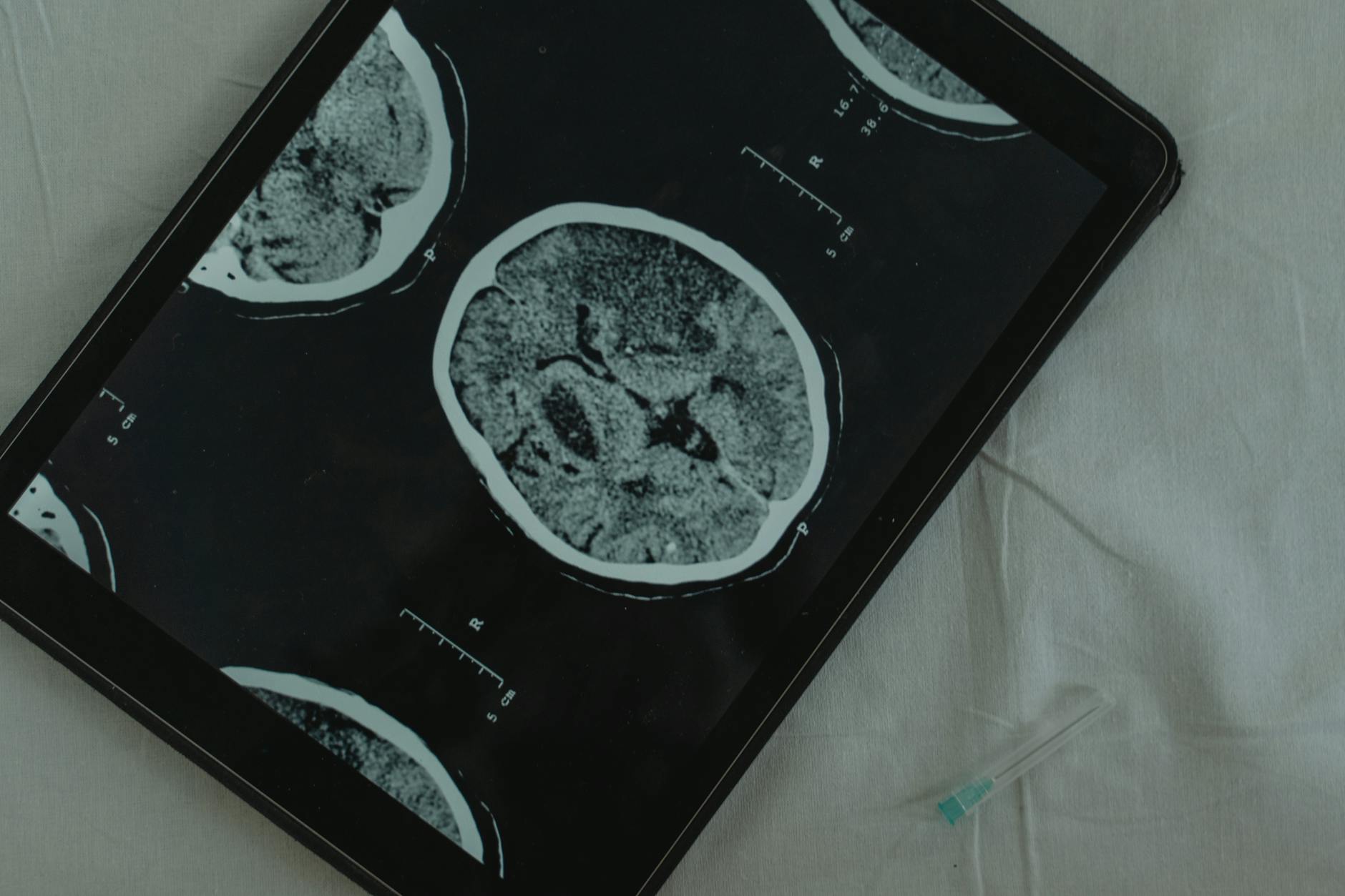
Brain scan results provide objective visual evidence of changes in the brain that may accompany dementia, helping doctors confirm what clinical assessments suggest and giving caregivers concrete information about what’s happening physically. When a loved one receives a dementia diagnosis, brain scans—typically MRI, PET, or sometimes both—can show structural shrinkage, abnormal protein buildup, or changes in how the brain uses glucose. For example, if your mother has been forgetting recent conversations and getting confused about familiar routes, an MRI might reveal significant shrinkage in her hippocampus, the brain region critical for forming new memories, while a PET scan could show diminished glucose metabolism in areas responsible for thinking and planning.
Understanding what these scans reveal helps caregivers grasp why their loved one is experiencing specific symptoms and prepares them for what may come next. Brain scans aren’t magic tools that diagnose dementia on their own—they must be interpreted alongside medical history, cognitive testing, and clinical evaluation—but they do provide measurable, visible confirmation that something in the brain has changed. Approximately 6.9 million Americans aged 65 and older currently live with Alzheimer’s disease, and many of their families benefit from the clarity that brain imaging provides.
Table of Contents
- What Do Different Brain Scans Show About Dementia?
- Understanding Amyloid and Tau on Brain Imaging—What the Biomarkers Mean
- How Brain Scans Help Confirm (or Rule Out) a Dementia Diagnosis
- What Caregivers Should Know When Reviewing Scan Results with the Doctor
- Brain Scans Can Miss Some Forms of Dementia—A Critical Limitation
- Advanced Imaging Options: When Standard Scans Aren’t Enough
- The Future of Brain Imaging in Dementia Care
- Conclusion
What Do Different Brain Scans Show About Dementia?
mri (magnetic resonance imaging) is typically the first brain scan ordered for suspected dementia because it clearly visualizes the brain’s structure. An MRI takes detailed pictures that reveal whether brain tissue has shrunk in key areas, particularly the hippocampus, which is often among the first regions affected by Alzheimer’s disease. Modern automated analysis of MRI scans achieves 90% sensitivity and 84% specificity in detecting dementia-related changes, meaning it correctly identifies most people with dementia while avoiding false alarms. When a radiologist reviews your loved one’s MRI, they’re looking for this characteristic pattern of shrinkage in specific regions, not just general brain size.
PET (positron emission tomography) scans work differently—they show how the brain is functioning, not just its structure. PET scans detect changes in glucose metabolism (how much energy different brain regions are using), the accumulation of amyloid and tau proteins, oxygen metabolism, and blood flow abnormalities. A person with dementia typically shows a distinctive pattern of abnormally decreased glucose use in specific brain areas, appearing as darker regions on the scan. While an MRI might show that the hippocampus is smaller, a PET scan reveals that cells throughout the frontal and temporal lobes are no longer working efficiently, burning less fuel than they should. Some medical centers now offer combined PET/MRI technology that captures both types of information in a single session, which can significantly reduce the inconvenience and cost for patients and caregivers who might otherwise need two separate appointments.
Understanding Amyloid and Tau on Brain Imaging—What the Biomarkers Mean
Amyloid and tau are proteins that accumulate abnormally in Alzheimer’s disease, and modern PET scans can detect them directly. The two proteins behave differently in the brain: amyloid tends to spread widely throughout the brain tissue, while tau protein concentrates precisely in the areas where brain atrophy is most severe. This distinction matters because it helps explain why two people with similar amyloid levels might have very different symptoms. The presence of both elevated amyloid and elevated tau biomarkers predicts the steepest cognitive decline over time, meaning caregivers should expect more rapid changes if their loved one tests positive for both.
One important limitation is that having abnormal biomarkers doesn’t guarantee someone will develop symptoms soon—some cognitively normal older adults show amyloid accumulation on scans but remain unaffected for years. Additionally, a single brain scan is a snapshot in time; biomarker progression happens at different rates for different people. If your loved one’s initial PET scan shows elevated tau, there’s a 93% likelihood that amyloid PET will also be positive, which can help guide treatment decisions but doesn’t predict exactly how quickly symptoms will progress. Brain scans provide valuable biological information, but they can’t tell you whether someone will develop symptoms next year or ten years from now.
How Brain Scans Help Confirm (or Rule Out) a Dementia Diagnosis
The 2024 revised diagnostic criteria for Alzheimer’s disease now formally incorporate biomarker findings, marking a significant shift toward objective, measurable diagnosis rather than relying solely on symptom assessment. This means that brain scans have become part of the medical standard for confirming dementia, especially when symptoms are subtle or when doctors need to distinguish Alzheimer’s from other conditions that can cause similar cognitive changes. For instance, if an elderly person is experiencing memory problems, an MRI might reveal that the brain changes are actually from small strokes or vascular damage rather than Alzheimer’s—a crucial distinction because treatment approaches differ significantly. However, brain scans cannot diagnose dementia on their own.
A radiologist cannot look at an MRI or PET scan and make a diagnosis without clinical context. Diagnosis requires the combination of brain imaging, neuropsychological testing (formal memory and thinking tests), medical history, physical examination, and observation of how the person actually functions in daily life. Someone might have scan abnormalities consistent with dementia yet perform normally on cognitive tests and have no functional decline—in that case, a diagnosis wouldn’t be made. The scan is one piece of a larger diagnostic puzzle, important but not sufficient by itself.
What Caregivers Should Know When Reviewing Scan Results with the Doctor
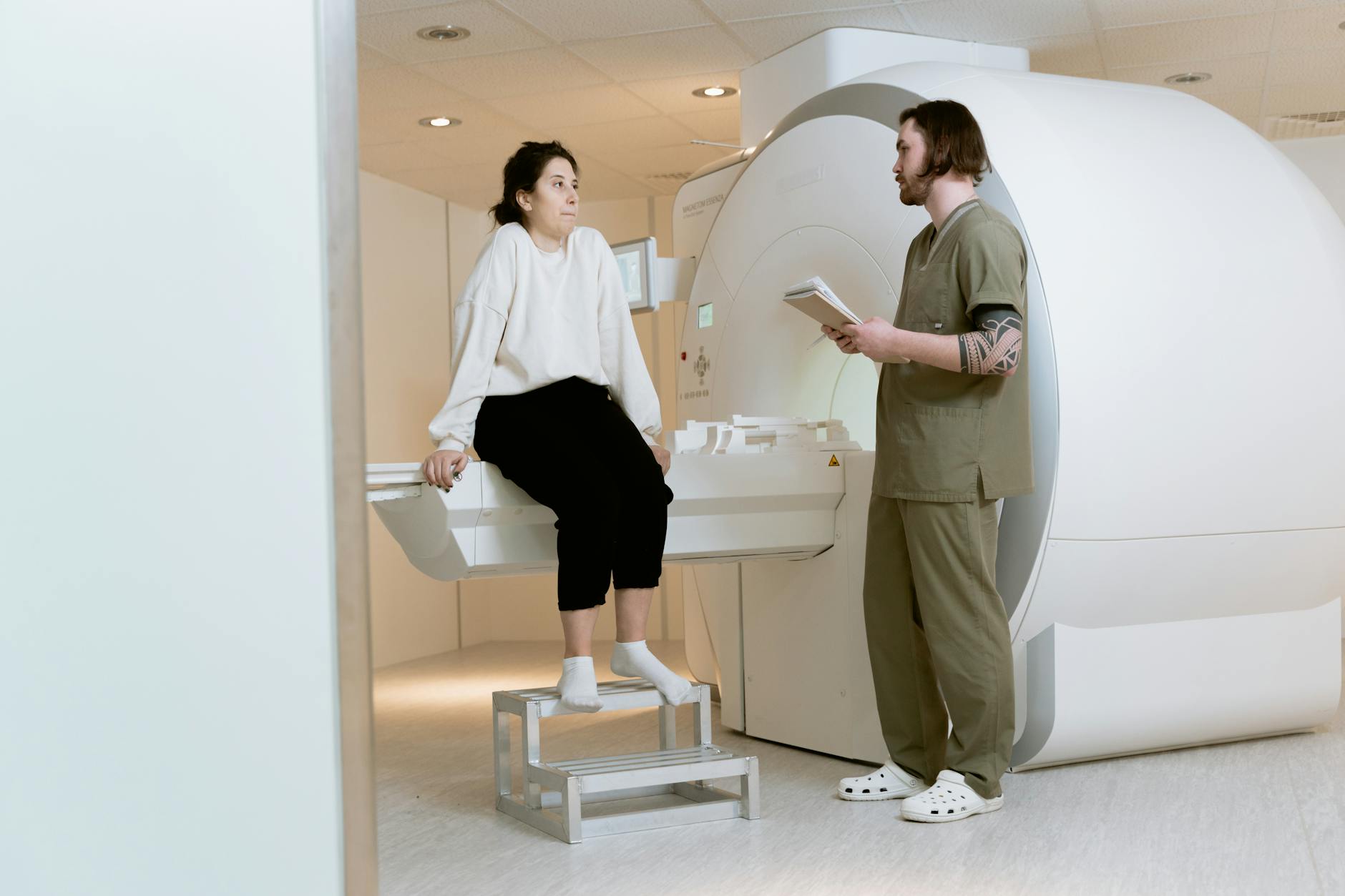
When your doctor presents brain scan results, ask specifically what structures show shrinkage, where biomarker abnormalities appear, and what those findings typically mean for progression and symptoms. A good explanation connects the scan findings to what you’ve already observed: “The MRI shows hippocampal atrophy, which explains why your husband is struggling with new information but still remembers events from years ago.” Understanding this connection helps you recognize which abilities might decline next and prepare accordingly. Write down the specific findings rather than relying on memory, so you can reference them during future appointments or when discussing your loved one’s care with family members. Be prepared for the possibility that scan results may be somewhat ambiguous.
Radiologists often note findings as “mild,” “moderate,” or “significant” rather than giving definitive yes-or-no answers. An MRI showing “some hippocampal atrophy” might be described in one person as consistent with normal aging and in another as consistent with early Alzheimer’s—the difference lies in how much atrophy is present and in the overall clinical context. If you don’t fully understand the explanation, ask the doctor to clarify or request written results. Some medical centers now offer combined PET/MRI reports that consolidate structural and functional information, which can be easier for families to understand than separate reports.
Brain Scans Can Miss Some Forms of Dementia—A Critical Limitation
While brain scans are powerful diagnostic tools, they have significant limitations that caregivers should understand. Some forms of dementia, particularly frontotemporal dementia and Lewy body dementia, may not show dramatic structural changes on MRI early in the disease course, even though cognitive decline is evident. A person might have clear behavioral changes, executive dysfunction, or hallucinations yet have a relatively normal-looking MRI. This is why doctors sometimes order additional scans or tests if the clinical picture doesn’t match the imaging findings.
Another critical warning: don’t assume that a “normal” brain scan rules out dementia. Mild cognitive impairment and early dementia may not yet produce visible changes on standard MRI. If your loved one shows cognitive symptoms but the brain scan looks relatively normal, the diagnosis isn’t automatically ruled out—additional testing, a repeat scan in several months, or specialist evaluation may be necessary. Conversely, some older adults have brain changes on scans that seem to warrant a dementia diagnosis, yet they function normally and have no actual cognitive decline. This disconnect between scan findings and real-world ability emphasizes that caregivers shouldn’t rely solely on what a scan shows; ongoing observation of your loved one’s actual functioning is equally important.

Advanced Imaging Options: When Standard Scans Aren’t Enough
Newer imaging techniques are emerging that can provide more detailed information than standard MRI and PET scans. High-field MRI (3T and 7T) offers better visualization of subtle changes and microstructural damage in white matter. Some specialized centers offer tau-specific PET tracers and amyloid-specific PET tracers that are more precise than older agents in mapping where these proteins accumulate. For example, a tau PET scan might reveal that tau is concentrated primarily in the medial temporal lobe, suggesting the diagnosis is likely Alzheimer’s disease rather than primary age-related tauopathy, which affects different brain regions.
Combined PET/MRI imaging is particularly valuable because it simultaneously captures both structural and functional information without requiring two separate procedures. For caregivers managing a loved one’s medical appointments, this consolidation reduces stress and costs. However, these advanced options aren’t universally available and may not be covered by insurance unless there’s a specific clinical indication. Discuss with your loved one’s neurologist whether these specialized scans would provide additional useful information for diagnosis or treatment planning in your specific situation.
The Future of Brain Imaging in Dementia Care
Biomarker research continues advancing, with new blood tests emerging that can detect amyloid, tau, and other disease markers from a simple blood draw, potentially reducing the need for PET scans in some situations. These blood biomarkers correlate well with imaging findings and may eventually allow earlier detection and intervention, before substantial brain changes occur. For caregivers, this shift means that future diagnostic pathways might be less invasive and more accessible, bringing high-quality dementia assessment to communities without specialized imaging centers.
Treatment approaches are also evolving in response to biomarker findings. Newer Alzheimer’s medications target amyloid and tau specifically, and knowing a person’s biomarker status—confirmed by brain scans or blood tests—helps doctors choose the most appropriate treatment. As imaging technology improves and biomarker understanding deepens, brain scans will likely play an even larger role in guiding personalized treatment decisions, making the information they provide increasingly relevant to your loved one’s actual care plan.
Conclusion
Brain scans offer caregivers valuable, objective information about the physical changes occurring in their loved one’s brain, but they should always be understood in context with clinical assessment, cognitive testing, and real-world observation of how the person is actually functioning. The combination of structural imaging (MRI) and functional imaging (PET) provides complementary information: MRI shows what has changed in brain structure, while PET reveals how well remaining brain tissue is working. Knowing what a scan shows—and just as importantly, knowing what it cannot show—helps you work more effectively with doctors and make informed decisions about care and treatment.
After receiving brain scan results, work with your doctor to understand what the findings mean for your loved one’s specific situation, ask how the scan results explain the symptoms you’ve observed, and discuss what to expect as the disease progresses. Keep detailed records of scan findings and dates so you can track changes over time. Remember that while brain scans provide important biological information, they’re just one tool among many in dementia diagnosis and care—your own careful observation of your loved one’s abilities, needs, and changes remains equally important to their medical management.