Reviewed by the Help Dementia Editorial Team — our editors review every article for accuracy against guidance from the National Institute on Aging, the Alzheimer’s Association, and peer-reviewed sources.
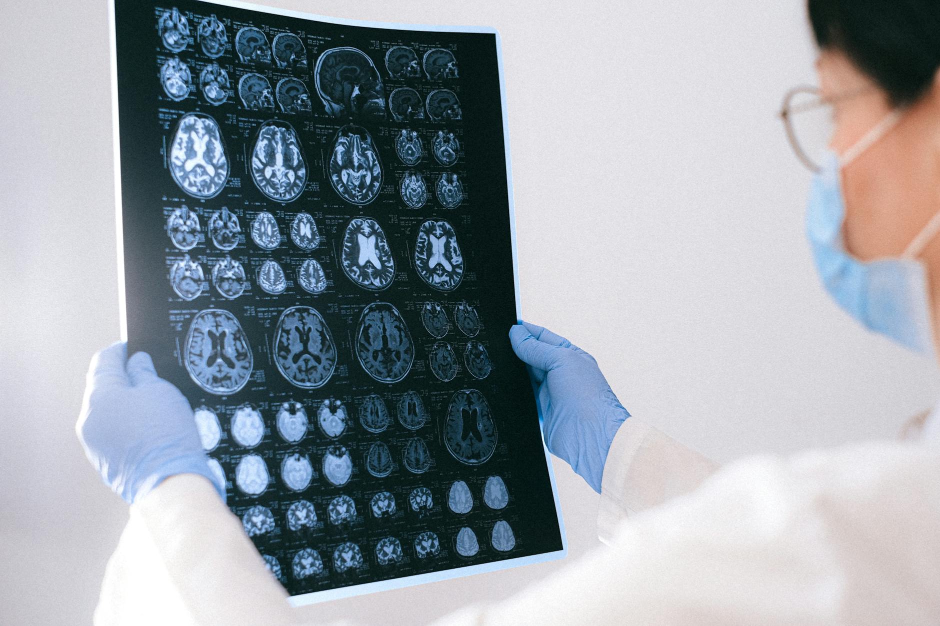
MRI scans can reveal significant changes in the brain structures of Alzheimer’s disease patients, particularly brain atrophy or shrinkage in specific regions like the temporal and parietal lobes. These structural changes become increasingly visible on MRI as the disease progresses, though early-stage Alzheimer’s may show a normal-appearing MRI despite ongoing cognitive decline.
The most characteristic finding is shrinkage of the hippocampus and medial temporal lobe, which researchers have identified as the hallmark MRI signature of Alzheimer’s disease. For example, a 68-year-old woman experiencing mild memory problems and difficulty with conversations might undergo an MRI that shows subtle hippocampal shrinkage—a finding that, combined with cognitive testing and symptom evaluation, helps physicians piece together whether Alzheimer’s disease is developing. However, it’s important to understand that MRI doesn’t provide a definitive diagnosis on its own; instead, it serves as one piece of evidence that, when combined with clinical evaluation and other factors, helps doctors understand what’s happening in the brain.
Table of Contents
- Brain Atrophy as the Key MRI Finding in Alzheimer’s Disease
- The Specific Brain Regions Affected by Alzheimer’s
- Four Distinct MRI-Detectable Atrophy Subtypes in Alzheimer’s Disease
- MRI’s Diagnostic Accuracy for Detecting Alzheimer’s Disease
- Important Limitations of MRI for Alzheimer’s Diagnosis
- Using MRI to Rule Out Other Conditions and Monitor Disease Progression
- The Future of MRI and Advanced Brain Imaging in Alzheimer’s Detection
- Conclusion
Brain Atrophy as the Key MRI Finding in Alzheimer’s Disease
brain atrophy refers to the loss of neurons and connections between them, which causes the brain tissue to shrink. In Alzheimer’s disease, this atrophy follows a predictable pattern, with the medial temporal lobe—a region deep inside the brain that includes the hippocampus—showing the most consistent changes. The hippocampus, which plays a central role in forming new memories, is typically the first region to show visible shrinkage on mri scans in Alzheimer’s patients. The distinction between early and late Alzheimer’s is important when interpreting MRI results.
In the early stages of the disease, a patient’s MRI might appear completely normal, even though cognitive decline has already begun. This counterintuitive finding—that brain structure looks fine on imaging even as memory and thinking are deteriorating—can sometimes delay diagnosis. As the disease progresses into moderate and later stages, the atrophy becomes more apparent and widespread, eventually affecting the temporal and parietal lobes more broadly. Understanding the timing of these changes helps explain why some people with mild cognitive impairment have normal MRIs: the pathological changes of Alzheimer’s are occurring at the cellular level before they become visible on imaging. This is why physicians may recommend MRI even when symptoms are subtle, as it can help establish a baseline and track changes over time.
The Specific Brain Regions Affected by Alzheimer’s
Research has identified that the medial temporal lobe atrophy is the hallmark MRI finding that distinguishes Alzheimer’s disease from other forms of dementia and normal aging. The hippocampus, located within this region, is particularly vulnerable because it contains high concentrations of amyloid plaques and tau tangles—the pathological hallmarks of Alzheimer’s—early in the disease process. When physicians or radiologists review an MRI of an Alzheimer’s patient, they’re looking for distinctive shrinkage or damage to this seahorse-shaped structure deep within the brain. Beyond the temporal lobe, Alzheimer’s typically progresses to affect the parietal lobes, which are responsible for spatial awareness, coordination, and processing sensory information.
This expanding pattern of atrophy helps distinguish Alzheimer’s from other conditions like frontotemporal dementia, which damages the front of the brain first, or vascular dementia, which may show patterns of small vessel disease or stroke-related changes instead of the characteristic atrophy pattern. One important limitation is that brain atrophy on MRI is not specific to Alzheimer’s disease alone. Age-related shrinkage, chronic conditions like diabetes or hypertension, and other types of dementia can also cause brain atrophy. This is why MRI findings must always be interpreted in context with a patient’s symptoms, cognitive testing results, medical history, and sometimes additional biomarker tests.
Four Distinct MRI-Detectable Atrophy Subtypes in Alzheimer’s Disease
Research has identified that Alzheimer’s disease doesn’t look identical in every patient’s brain. Scientists have distinguished four different MRI-detectable atrophy subtypes within the Alzheimer’s disease population: typical AD, limbic-predominant AD, hippocampal-sparing AD, and minimal atrophy subtype. Understanding these subtypes is helping researchers and clinicians recognize that Alzheimer’s disease may present with varying patterns of brain damage, even though the underlying pathology—amyloid and tau accumulation—is fundamentally the same. The typical AD subtype shows the pattern most commonly described in textbooks: prominent medial temporal and parietal lobe atrophy. The limbic-predominant subtype shows greater atrophy in areas involved in emotion and memory regulation.
The hippocampal-sparing subtype, as its name suggests, shows less damage to the hippocampus but more atrophy in other cortical regions. The minimal atrophy subtype shows relatively little structural change on MRI despite cognitive decline. These distinctions matter because different atrophy patterns may correlate with variations in how quickly the disease progresses and which symptoms emerge first. For instance, someone with hippocampal-sparing Alzheimer’s might experience language difficulties or executive function problems earlier than memory loss, since their brain damage is concentrated in regions that support language and planning rather than memory formation. Identifying these subtypes helps explain why Alzheimer’s disease presents differently from person to person and why treatment approaches may need to be tailored to individual patterns of brain damage.
MRI’s Diagnostic Accuracy for Detecting Alzheimer’s Disease
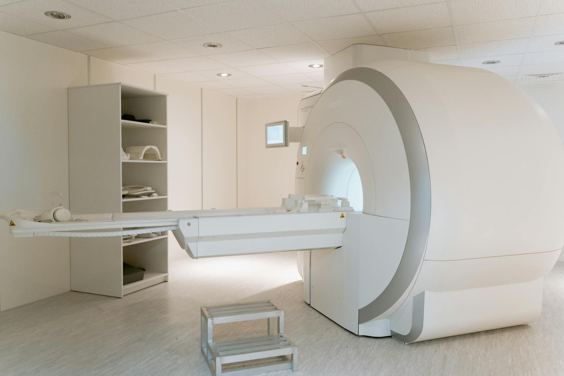
MRI has demonstrated remarkable diagnostic accuracy for identifying Alzheimer’s disease, particularly in early stages before symptoms become obvious. Studies have shown that MRI can achieve up to 87 percent diagnostic accuracy for confirming Alzheimer’s disease up to seven years before symptom onset—meaning the imaging can detect brain changes associated with the disease years before a person notices memory problems or cognitive decline. One particularly rigorous study achieved 89.1 percent sensitivity and 87.0 percent specificity, which exceeded the 85 percent performance level of radiologists analyzing images without additional patient information. These accuracy rates represent a significant advance in diagnostic capability.
For comparison, clinical diagnosis based on symptoms alone is often less reliable, particularly in mild stages of cognitive impairment where symptoms may be subtle or overlap with normal aging. MRI can identify which patients with mild cognitive impairment are likely to develop Alzheimer’s disease, providing both patients and their families with prognostic information and an opportunity to plan for the future. However, these impressive statistics come with an important caveat: they represent research findings from specialized centers with experienced radiologists and dedicated research protocols. In routine clinical practice, the diagnostic accuracy may be somewhat lower, and MRI findings are most valuable when combined with cognitive testing, symptom assessment, and sometimes additional biomarker tests like blood work for phosphorylated tau or amyloid levels.
Important Limitations of MRI for Alzheimer’s Diagnosis
While MRI is a valuable tool, it has important limitations that every patient and family member should understand. Most critically, no single test, including MRI, can definitively diagnose Alzheimer’s disease on its own. Even with the high diagnostic accuracy rates mentioned above, MRI findings must be interpreted as part of a comprehensive clinical evaluation that includes assessment of cognitive symptoms, medical history, and exclusion of other conditions that can cause dementia. In early-stage Alzheimer’s disease, MRI may appear completely normal despite significant pathological changes occurring in the brain at the cellular level.
This means that a normal MRI does not rule out Alzheimer’s disease; it simply means that visible structural changes aren’t yet apparent on this particular imaging modality. Some patients with cognitive decline may have both Alzheimer’s pathology and other contributing factors like vascular changes, Lewy body disease, or frontotemporal changes, resulting in a more complex clinical picture than MRI alone can clarify. Additionally, MRI is not sensitive enough to detect the earliest molecular changes of Alzheimer’s disease. The amyloid plaques and tau tangles that define Alzheimer’s pathology at the microscopic level cannot be visualized on standard structural MRI. This is why researchers increasingly use specialized imaging like PET scans for amyloid and tau, though these advanced imaging techniques are typically reserved for research settings or specialized dementia clinics rather than routine clinical practice.

Using MRI to Rule Out Other Conditions and Monitor Disease Progression
One of MRI’s most practical applications in dementia evaluation is its ability to exclude other causes of cognitive decline. A patient experiencing memory problems might actually have a treatable condition like normal pressure hydrocephalus, a brain tumor, or previous strokes—all of which show characteristic patterns on MRI. By identifying these alternative diagnoses, MRI prevents unnecessary treatment delays and directs patients toward appropriate therapy.
MRI also plays an important role in longitudinal monitoring, allowing physicians to track brain changes over time. Serial MRI scans—taken months or years apart—can document whether brain atrophy is progressing as expected, remaining stable, or showing unexpected changes that might suggest a different diagnosis or complication. This monitoring function helps assess how someone is responding to treatments and whether cognitive decline is tracking with structural brain changes.
The Future of MRI and Advanced Brain Imaging in Alzheimer’s Detection
As our understanding of Alzheimer’s disease advances, so too are imaging technologies evolving. High-field MRI scanners and specialized sequences are becoming increasingly sensitive to subtle brain changes, potentially allowing earlier detection of the disease process.
Researchers are also developing ways to combine MRI findings with blood biomarkers—such as phosphorylated tau and amyloid levels—to create more comprehensive diagnostic profiles that don’t rely on any single test. The combination of structural MRI, functional imaging, and molecular biomarkers represents the likely future of Alzheimer’s diagnosis, moving away from reliance on any single test toward a multi-modal assessment approach. This evolution promises to make early detection and diagnosis increasingly accurate and accessible.
Conclusion
MRI reveals characteristic brain atrophy patterns in Alzheimer’s disease, most notably shrinkage of the medial temporal lobe and hippocampus, with four distinct atrophy subtypes now recognized by researchers. These structural changes can sometimes be detected years before symptoms appear, and MRI has demonstrated up to 87 percent diagnostic accuracy when used appropriately—though early-stage disease may show normal imaging despite ongoing pathological changes.
However, MRI must always be interpreted as part of a comprehensive clinical evaluation that includes cognitive testing, symptom assessment, and consideration of alternative diagnoses. If you or a family member is experiencing cognitive concerns, MRI may be one component of a thorough evaluation, but should be combined with medical history review, cognitive screening, and discussion with a healthcare provider familiar with dementia evaluation to establish an accurate diagnosis and appropriate care plan.