Reviewed by the Help Dementia Editorial Team — our editors review every article for accuracy against guidance from the National Institute on Aging, the Alzheimer’s Association, and peer-reviewed sources.
Brain atrophy and dementia are related but fundamentally different conditions. Brain atrophy is the physical loss of brain tissue, cells, and neural connections that leads to a measurable decrease in brain volume. Dementia, by contrast, is a clinical syndrome—a progressive decline in cognitive function that affects a person’s ability to think, remember, communicate, and live independently. A person can experience brain atrophy without developing dementia, just as some people with dementia may not show severe brain shrinkage on imaging. The key distinction is that atrophy is a structural change you can see on a brain scan, while dementia is a functional decline you experience in daily life.
Consider Margaret, a 72-year-old woman whose MRI revealed moderate hippocampal atrophy—the brain region critical for memory. Her doctor explained that while this finding was concerning, it didn’t automatically mean she had dementia. Margaret still remembered appointments, managed her finances, and lived independently. However, she was at elevated risk, so her doctor recommended regular cognitive screening and lifestyle interventions to slow progression. This distinction matters because it shapes how we approach treatment, monitoring, and expectations.
Table of Contents
- How Does Brain Atrophy Differ from Dementia?
- Understanding the Brain Regions Most Affected by Atrophy
- What Modern Brain Imaging Reveals About Atrophy and Dementia
- The Relationship Between Brain Atrophy and Dementia Risk
- When Brain Atrophy Progresses Without Obvious Cognitive Symptoms
- How Hippocampal Atrophy Predicts Progression to Dementia
- Living with Brain Atrophy: Monitoring, Prevention, and Hope
- Conclusion
How Does Brain Atrophy Differ from Dementia?
brain atrophy involves the death or loss of neurons and the connections between them, resulting in shrinkage of brain tissue. This can be measured on advanced MRI scans, which show reduced volume in specific brain regions. Some degree of brain atrophy is normal with aging—the brain naturally loses about 5% of its volume per decade after age 40. However, the rate and location of atrophy matter significantly. In Alzheimer’s disease, atrophy in the hippocampus occurs at a rate of 4.66% per year, compared to just 1.41% annually in healthy older adults. This accelerated loss indicates pathological change.
Dementia, meanwhile, is diagnosed based on cognitive and functional changes, not brain scans. A person has dementia when cognitive decline interferes with daily activities—forgetting important events repeatedly, getting lost in familiar places, struggling to manage finances or medications, or difficulty with language. Dementia is what someone experiences and how it affects their life; atrophy is what’s visible on neuroimaging. Many people with brain atrophy never develop noticeable cognitive symptoms, especially if the atrophy is mild or occurs in non-critical regions. The relationship is indirect but real: severe brain atrophy in specific regions—particularly the hippocampus, temporal cortex, and amygdala—significantly increases the risk of developing dementia. Studies tracking thousands of older adults show that hippocampal atrophy is the strongest structural predictor of who will progress to Alzheimer’s dementia in the coming years. But a single MRI snapshot doesn’t diagnose dementia; it identifies risk.

Understanding the Brain Regions Most Affected by Atrophy
When dementia develops, brain atrophy doesn’t occur uniformly. The damage concentrates in specific regions critical for memory, emotion, and language. The hippocampus—a seahorse-shaped structure deep in the brain—is typically the first and hardest hit in Alzheimer’s disease. The temporal cortex, which processes memories and language, also shrinks substantially. The amygdala, which handles emotional processing, and the temporal lobes more broadly show consistent volume loss in dementia patients compared to healthy controls. Meta-analyses combining data from hundreds of research participants reveal a clear pattern: the more severe the dementia, the greater the volume loss across multiple brain regions.
In one major analysis of 595 Alzheimer’s patients and 212 matched controls across nine studies, hippocampal atrophy measured on MRI showed strong correlation with disease severity. Importantly, the fastest hippocampal volume loss occurs in people with active Alzheimer’s disease compared to normal aging in cognitively intact older adults. This rapid deterioration is what distinguishes pathological atrophy from normal age-related change. However, there’s an important limitation: brain volume loss doesn’t always correlate perfectly with cognitive symptoms. Two people with similar amounts of atrophy may experience different levels of functional decline. This highlights that brain structure, cognitive reserve, brain plasticity, and individual resilience all influence whether someone develops dementia or how quickly symptoms progress.
What Modern Brain Imaging Reveals About Atrophy and Dementia
Advanced MRI technology can now detect brain atrophy with remarkable precision. Automated volumetry—computer-assisted measurement of brain structures—shows 90% sensitivity and 84% specificity in identifying dementia-related changes. High-field 3T MRI scanners and ultra-high-field 7T systems can detect very subtle structural changes that older imaging equipment would miss. These advances allow doctors to identify people at high risk for progression years before cognitive symptoms become obvious. However, MRI findings are not diagnostic for dementia on their own.
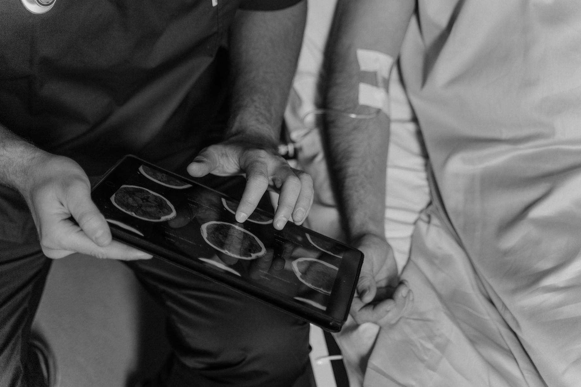
Many cognitively normal older adults show some degree of atrophy on imaging without any functional decline. Conversely, some people with cognitive symptoms may have relatively preserved brain volume. The imaging must be interpreted in context with cognitive testing, functional status, and medical history. This is a critical limitation: seeing atrophy on a brain scan should prompt careful evaluation and monitoring, but it doesn’t automatically mean someone has dementia or will develop it soon. The practical value of these scans is in tracking change over time. A single MRI provides a snapshot, but serial imaging—comparing scans taken months or years apart—reveals the rate of atrophy and helps predict who is likely to progress.
The Relationship Between Brain Atrophy and Dementia Risk
The statistical relationship between atrophy and dementia risk is substantial but not deterministic. Current prevalence data shows that 7.2 million Americans age 65 and older live with Alzheimer’s dementia today, with rates increasing sharply with age: 5% of those 65-74 have Alzheimer’s, compared to 13% of those 75-84, and 33% of those age 85 and older. Without major medical advances, this number is projected to grow to 13.8 million by 2060. Perhaps most striking is a 2025 estimate that people have a 42% lifetime risk of developing dementia after age 55—more than double previous estimates.
This means that for many people, brain changes are accumulating throughout their 60s, 70s, and 80s, potentially including atrophy, even if symptoms haven’t yet appeared. The implication is that brain atrophy in middle-aged and older adults should be taken seriously as a sign of elevated future risk, not dismissed as inconsequential. The practical tradeoff is between reassurance and vigilance. Learning that you have brain atrophy on an MRI can be frightening, but it doesn’t automatically mean you’ll develop dementia. The key is understanding your individual risk factors and adopting protective strategies now—cardiovascular health, cognitive engagement, physical activity, quality sleep, social connection, and Mediterranean-style diet are all linked to slowing atrophy progression.
When Brain Atrophy Progresses Without Obvious Cognitive Symptoms
One of the most challenging scenarios in neurology is the person with clear evidence of brain atrophy on imaging who performs normally on cognitive testing. This situation can persist for years, creating uncertainty and anxiety. The person knows structural damage is occurring but isn’t experiencing the functional consequences—yet. This discrepancy reflects the concept of “cognitive reserve,” the idea that people vary in their ability to tolerate brain damage before symptoms emerge. A critical warning: normal cognitive testing at one point doesn’t guarantee that atrophy won’t cause problems later.
The same structural damage that’s asymptomatic today may cross a threshold in months or years and suddenly manifest as memory loss, confusion, or language difficulty. This is why people with documented brain atrophy benefit from regular cognitive screening—not yearly, but every few years—to detect the earliest signs of functional decline before they interfere with independence. Additionally, people sometimes ask whether they should have multiple MRI scans to track atrophy progression. While serial imaging can be valuable, doing too many scans exposes you to unnecessary cumulative cost and, with some contrast agents, potential risks. The recommendation is typically to rescan 12-24 months after an initial abnormal finding, rather than annually.

How Hippocampal Atrophy Predicts Progression to Dementia
The hippocampus is so central to memory that its atrophy in older adults deserves specific attention. Research shows that people with established Alzheimer’s disease lose hippocampal volume at roughly 3.5 to 4.66% per year, compared to 1.4% per year in healthy controls. That threefold difference is substantial and detectable on serial MRI scans separated by just 12 months.
More importantly, hippocampal atrophy predicts who will transition from normal cognition to mild cognitive impairment, and eventually to dementia. Someone with significant hippocampal volume loss and normal cognition today has a much higher probability of developing cognitive symptoms within the next 2-5 years than someone with preserved hippocampal volume. This makes hippocampal measurement one of the most clinically useful markers for predicting dementia risk, more informative than general brain atrophy in other regions.
Living with Brain Atrophy: Monitoring, Prevention, and Hope
For people diagnosed with brain atrophy, the path forward involves three components: understanding what’s happening, monitoring progression carefully, and implementing evidence-based strategies to slow decline. This is not a fate but a manageable condition requiring attention and action. Mediterranean diet adherence, aerobic exercise, cognitive stimulation, quality sleep, hearing correction if needed, and social engagement all have evidence supporting their role in slowing cognitive decline.
The landscape is also shifting toward earlier intervention. New anti-amyloid monoclonal antibodies like lecanemab have shown modest benefit in slowing cognitive decline in early Alzheimer’s disease, potentially offering a small window of opportunity for people with atrophy and very early cognitive changes. While these drugs are not cures, they represent the first disease-modifying treatments that can slow progression, offering hope that future treatments will be more effective.
Conclusion
Brain atrophy and dementia are distinct but interconnected: atrophy is a structural change visible on imaging, while dementia is a functional syndrome affecting daily life. You can have atrophy without dementia, though significant atrophy in critical brain regions—especially the hippocampus—increases the risk of developing cognitive decline. Current statistics show 7.2 million Americans with Alzheimer’s dementia today, with rates highest in those over 85, and a 42% lifetime risk of dementia after age 55.
If you or a loved one has been told about brain atrophy on an MRI, the appropriate response is informed monitoring and proactive health management, not resignation. Work with your neurologist or primary care doctor to establish a baseline, schedule appropriate follow-up imaging, perform regular cognitive screening, and adopt lifestyle strategies proven to support brain health. Understanding the difference between structural change and functional decline empowers you to make decisions and take actions that matter.