Reviewed by the Help Dementia Editorial Team — our editors review every article for accuracy against guidance from the National Institute on Aging, the Alzheimer’s Association, and peer-reviewed sources.
Mri show sits at the center of this dementia and brain health question.
An MRI can reveal structural changes in the brain that are often associated with dementia, but it cannot definitively diagnose dementia on its own. An MRI might show brain atrophy, white matter changes, or other abnormalities that suggest dementia, but a diagnosis requires a combination of imaging results, cognitive testing, medical history, and clinical evaluation. For example, an MRI might show significant shrinkage in the hippocampus, a brain region critical for memory, which is consistent with Alzheimer’s disease, yet that same finding could appear in a person with normal cognitive function or be caused by other conditions entirely.
MRI is one tool in the diagnostic toolkit for dementia evaluation, but it functions as supporting evidence rather than definitive proof. The technology excels at ruling out other conditions that mimic dementia symptoms—like brain tumors, stroke, or normal pressure hydrocephalus—which can be treated differently. When combined with neuropsychological testing, clinical interviews, and sometimes other imaging or biomarker tests, MRI helps doctors narrow the possibilities and support their clinical judgment.
Table of Contents
- How Does MRI Help Identify Brain Changes Related to Dementia?
- What Limitations Should You Know About MRI in Dementia Diagnosis?
- What Brain Changes Does an MRI Actually Show in Dementia Patients?
- How Does MRI Compare to Other Diagnostic Tools for Dementia?
- What Are the Risks and Misconceptions About MRI for Dementia Diagnosis?
- When Is MRI Most Useful in Dementia Evaluation?
- The Future of Brain Imaging in Dementia Diagnosis
- Conclusion
- Frequently Asked Questions
How Does MRI Help Identify Brain Changes Related to Dementia?
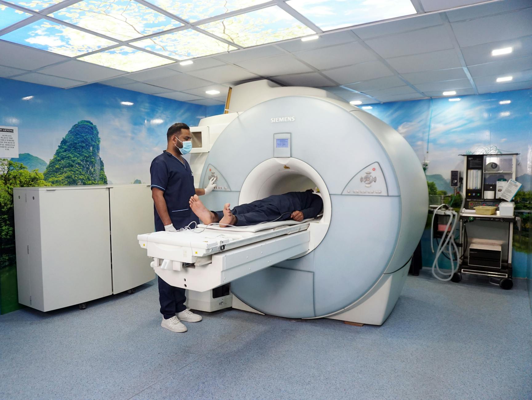
MRI creates detailed images of brain structures by using magnetic fields and radio waves, allowing doctors to visualize atrophy, tissue changes, and patterns of damage that often accompany dementia. The technology is particularly useful for identifying hippocampal shrinkage, which is a hallmark of Alzheimer’s disease, and for spotting vascular changes that suggest vascular dementia—the kind caused by reduced blood flow to the brain. However, some people show significant brain atrophy on MRI yet remain cognitively intact, while others with minimal visible changes on imaging may have severe cognitive decline, demonstrating that the relationship between what the MRI shows and actual cognitive function is not always straightforward. Different types of dementia produce different patterns on MRI.
In Alzheimer’s disease, doctors typically look for atrophy in the medial temporal lobe. In frontotemporal dementia, they watch for changes in the frontal and temporal lobes. Lewy body dementia may show relatively mild changes on conventional MRI. A radiologist reviewing these images has learned to recognize patterns, but those patterns alone do not make a dementia diagnosis—they contribute to the larger clinical picture.
What Limitations Should You Know About MRI in Dementia Diagnosis?
One critical limitation is that brain changes visible on MRI are not specific to dementia. Normal aging produces brain atrophy in many people who never develop cognitive problems. Some individuals in their 70s and 80s show significant atrophy and white matter hyperintensities (bright spots on the image indicating tissue changes) but maintain sharp minds. This creates a genuine challenge: doctors must distinguish between normal aging and pathological change, and an MRI alone cannot make that distinction.
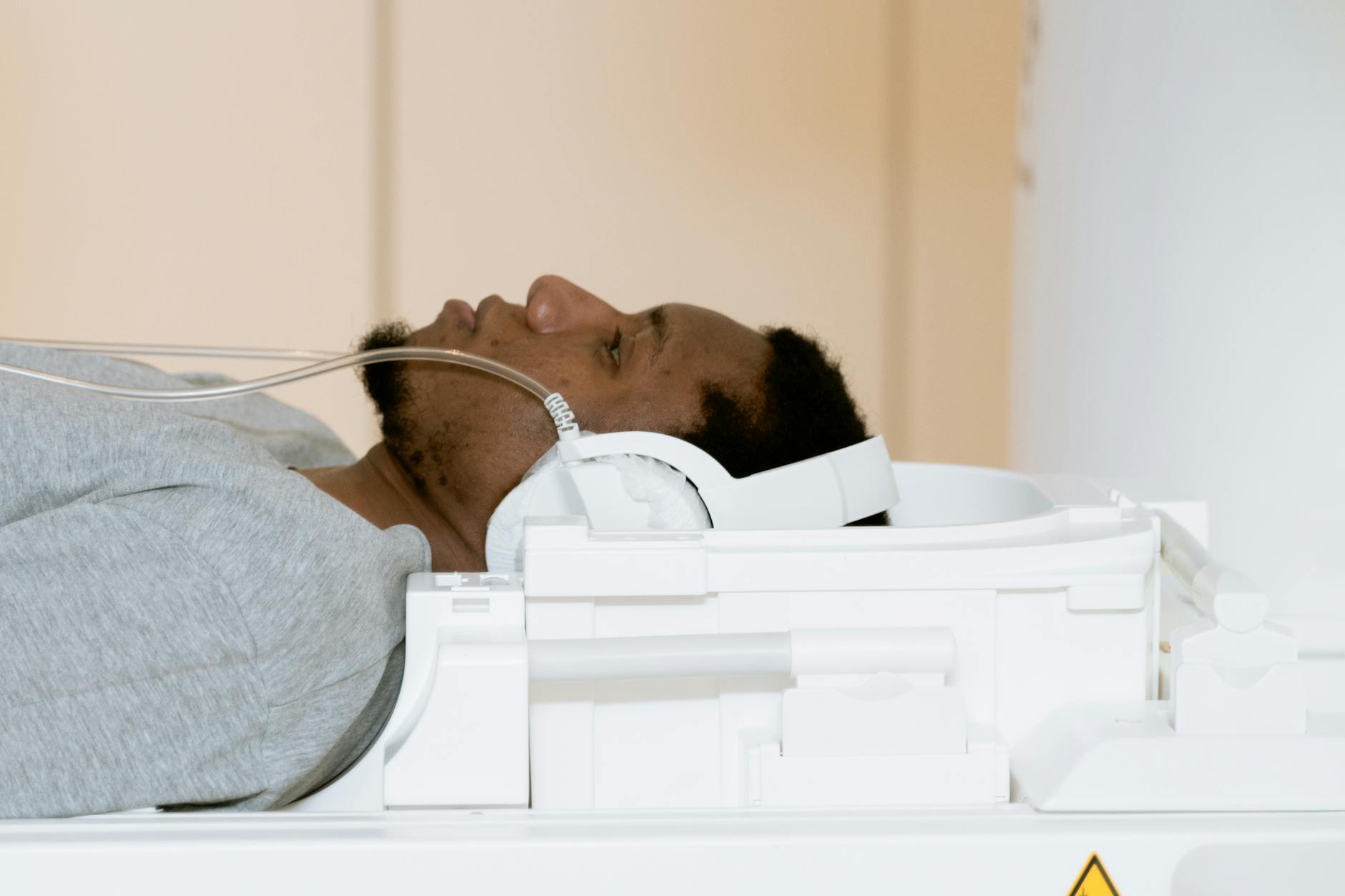
Another limitation is that early-stage dementia, particularly early Alzheimer’s disease, may not produce obvious changes visible on a standard MRI. Some people with confirmed Alzheimer’s pathology on biomarker testing show minimal structural changes on conventional imaging. Additionally, MRI is expensive, requires the patient to remain still for 30 to 60 minutes in a loud machine, and is not suitable for people with certain implants like older pacemakers or some metal hardware. For patients with claustrophobia or anxiety, the test can be difficult or impossible to complete. Insurance may not cover MRI for dementia evaluation unless specific clinical criteria are met, placing financial barriers in front of families seeking answers.
What Brain Changes Does an MRI Actually Show in Dementia Patients?
An MRI can visualize several structural changes associated with dementia: hippocampal atrophy (shrinkage of the memory-processing center), cortical atrophy (thinning of the brain’s outer layer), white matter hyperintensities (areas of damaged tissue), and ventricular enlargement (expansion of fluid-filled spaces in the brain). In vascular dementia, the MRI often reveals multiple infarcts—small areas of dead brain tissue caused by blocked blood vessels—sometimes creating a pattern that clinicians recognize. A patient presenting with memory loss, difficulty with words, and a family history of dementia might receive an MRI showing marked hippocampal atrophy, which substantially supports an Alzheimer’s diagnosis, especially when combined with positive cognitive testing results.
The specificity of these findings varies. Hippocampal atrophy is reasonably specific to Alzheimer’s disease compared to other dementias, but it appears in other conditions too, including depression, post-traumatic stress disorder, and even chronic stress in healthy individuals. White matter hyperintensities are extremely common in aging and can reflect vascular disease, but they also occur in people without cognitive decline. Radiologists measure atrophy using various rating scales and comparison to age-matched controls, but these measurements contain inherent subjectivity and variability between different radiologists’ assessments.
How Does MRI Compare to Other Diagnostic Tools for Dementia?
Cognitive testing, such as the Mini-Cog or Montreal Cognitive Assessment, directly evaluates memory, attention, language, and problem-solving—the actual abilities that matter clinically—whereas MRI shows only brain structure. Many neurologists prioritize cognitive testing and clinical evaluation over imaging in the initial diagnostic process. PET imaging, another neuroimaging technology, can detect abnormal protein accumulation in the Alzheimer’s brain before structural changes appear on MRI, making it more sensitive for early disease but also more expensive and involving radiation exposure.
Blood biomarkers—tests that measure levels of tau, phosphorylated tau, and amyloid beta in the bloodstream—are emerging as sensitive early indicators of Alzheimer’s pathology and are increasingly available through standard blood draws, offering advantages over imaging in terms of cost, accessibility, and early detection. The tradeoff is that while MRI provides structural detail that can rule out other diagnoses and identify patterns, it does not measure cognitive function directly. A neuropsychologist administering cognitive tests can identify the specific domains affected, such as memory versus language versus visuospatial skills, which helps differentiate between types of dementia. Many modern diagnostic approaches use MRI as one piece of evidence alongside cognitive testing, biomarkers, and clinical history rather than relying on imaging as the primary diagnostic tool.
What Are the Risks and Misconceptions About MRI for Dementia Diagnosis?
A significant misconception is that an MRI can definitively rule in or rule out dementia. Families sometimes believe that a “normal” MRI means no dementia is present, but absence of visible atrophy does not exclude dementia, particularly in early stages. Conversely, an MRI showing atrophy does not guarantee a dementia diagnosis—the findings must be interpreted in the context of the patient’s actual cognitive symptoms and test results. This gap between imaging findings and clinical reality creates confusion and sometimes false reassurance or unnecessary alarm.
Another consideration is overreliance on MRI results without adequate cognitive evaluation. A patient with memory complaints might receive an MRI that shows age-appropriate changes, after which the doctor concludes “the imaging looks normal, there’s nothing wrong.” This can delay diagnosis of actual dementia and deprive the patient of early intervention. Alternatively, when an MRI reveals atrophy but cognitive testing is not thorough, patients may be diagnosed with dementia based primarily on imaging, potentially labeling cognitively intact individuals or those with mild cognitive impairment as demented. The technology is powerful but requires skilled interpretation within a comprehensive clinical framework.

When Is MRI Most Useful in Dementia Evaluation?
MRI is particularly valuable when a patient’s symptoms could stem from a treatable condition that mimics dementia. A person experiencing memory loss and confusion might actually have a brain tumor, subdural hematoma, or normal pressure hydrocephalus—all of which require different treatment. MRI can identify these conditions and redirect the clinical course.
It is also useful when the clinical presentation is atypical or rapidly progressive, as these scenarios warrant investigation for alternative diagnoses. MRI is also part of the workup when vascular dementia is suspected, since the imaging can reveal the pattern and extent of vascular damage. For research purposes and specialized dementia centers, MRI combined with biomarker testing helps confirm the underlying pathology and guides enrollment into clinical trials. In these contexts, MRI provides concrete structural evidence that informs both clinical management and research advancement.
The Future of Brain Imaging in Dementia Diagnosis
Advanced imaging techniques are expanding the role of neuroimaging in dementia diagnosis. Amyloid and tau PET imaging, though currently expensive and not widely available, can detect the pathological proteins of Alzheimer’s disease before symptoms appear, potentially enabling intervention at earlier stages. MRI technology is also improving with newer sequences that can detect microstructural changes and subtle white matter damage earlier in the disease process.
Blood biomarkers are progressing rapidly and may eventually replace some imaging in routine clinical practice due to their accessibility and cost-effectiveness. The trajectory suggests a future in which dementia diagnosis integrates multiple modalities—cognitive testing, blood biomarkers, advanced structural and molecular imaging, and genetic risk assessment—to create a more complete picture. Standard MRI will likely remain part of this toolkit, particularly for ruling out structural mimics and providing supporting evidence, but it will be one component among several rather than the central diagnostic test.
Conclusion
An MRI can show brain changes associated with dementia, but it cannot diagnose dementia on its own. The images may reveal atrophy, white matter changes, or other abnormalities that fit with a dementia diagnosis, yet these findings require interpretation within the context of cognitive testing, clinical history, and patient symptoms. MRI’s greatest strength is ruling out other conditions that cause dementia-like symptoms and supporting clinical judgment when combined with other diagnostic tools.
If you or a family member is experiencing cognitive changes, seeking evaluation from a neurologist, neuropsychologist, or geriatrician is essential. These specialists can order appropriate testing, including MRI if indicated, interpret the results correctly, and develop a diagnostic and treatment plan tailored to the individual. Early and accurate diagnosis, supported by comprehensive evaluation rather than imaging alone, provides the best foundation for planning care and exploring treatment options.
Frequently Asked Questions
Does a normal MRI mean I don’t have dementia?
Not necessarily. A normal MRI does not exclude dementia, especially in early stages when brain changes may not be visible on standard imaging. Dementia diagnosis requires cognitive testing and clinical evaluation, not imaging alone.
Can an MRI show Alzheimer’s disease specifically?
An MRI can show patterns, like hippocampal atrophy, that are consistent with Alzheimer’s disease and more common in Alzheimer’s than other dementias. However, the same findings can appear in other conditions, so imaging alone cannot definitively confirm Alzheimer’s disease.
Is an MRI always needed when someone has dementia symptoms?
No. If cognitive testing and clinical evaluation clearly indicate dementia and there are no signs of other conditions, an MRI may not be necessary. Doctors typically order MRI when the presentation is unusual, rapidly progressive, or when other diagnoses need to be ruled out.
How much does an MRI cost for dementia evaluation?
MRI costs vary widely by location and facility, typically ranging from $500 to $3,000, often higher without insurance. Insurance coverage depends on whether the MRI meets specific medical necessity criteria established by the insurer.
What happens if the MRI is inconclusive?
If the MRI shows changes that could fit multiple diagnoses or shows minimal changes despite cognitive symptoms, additional testing such as cognitive neuropsychological testing, biomarker blood tests, or specialist consultation may help clarify the diagnosis.
Should I get an MRI if I’m worried about dementia?
Discuss your concerns with your doctor. They can assess whether MRI is appropriate based on your symptoms, medical history, and cognitive screening results. Often, cognitive testing occurs first to determine whether further imaging is warranted.
You Might Also Like
- How Long Can Someone Live With Vascular Dementia?
- How Long Can Someone Live With Dementia?
- MMSE Score of 20: Is It a Sign of Dementia?
For more, see NIH MedlinePlus — cognitive testing.