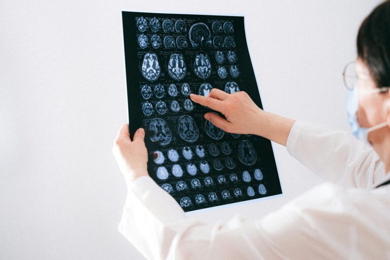
When a brain scan report mentions hippocampal atrophy, it means the hippocampus — a curved, seahorse-shaped structure deep in the temporal lobe — has lost tissue and shrunk compared to what would be expected for a person’s age. The hippocampus is the brain’s central hub for forming new memories and navigating space, and when it shrinks, those functions begin to suffer. Hippocampal atrophy is the most established biomarker of neurodegeneration that radiologists can detect on an MRI scan, visible as reduced volume, increased fluid space around the structure, and changes to the architecture of the medial temporal lobe. Seeing this finding on a scan report is understandably alarming, but the meaning depends heavily on degree, rate of change, and clinical context.
Not all hippocampal atrophy signals disease. Some shrinkage is a normal part of aging — research shows the hippocampus loses roughly 1.4% of its volume per year in healthy older adults. The concern arises when that rate accelerates significantly, or when atrophy appears at a degree inconsistent with a person’s age. In Alzheimer’s disease, the annual volume loss averages around 4.66%, more than three times the normal rate. This article covers what the finding means structurally, which conditions are associated with it, what the numbers actually indicate, and what recent research is revealing about detecting atrophy even earlier than current scans allow.
Table of Contents
- What Does the Hippocampus Do, and What Does Atrophy Look Like on a Brain Scan?
- How Much Atrophy Is Too Much — Understanding the Numbers
- Which Conditions Are Associated With Hippocampal Atrophy?
- What Happens After a Radiologist Flags Hippocampal Atrophy?
- What the Finding Does Not Mean — Important Limitations and Caveats
- What Recent Research Reveals About Earlier Detection
- The Future of Hippocampal Imaging in Dementia Medicine
- Conclusion
- Frequently Asked Questions
What Does the Hippocampus Do, and What Does Atrophy Look Like on a Brain Scan?
The hippocampus sits in the medial temporal lobe on both sides of the brain. Its primary roles include encoding new declarative memories — the kind that allow you to remember a conversation from last week or learn a new route — and supporting spatial navigation. Damage to the hippocampus, even before significant shrinkage is visible, begins to impair these functions. The memory problems that define early Alzheimer’s disease, particularly difficulty retaining new information while older memories stay relatively intact, reflect the hippocampus being one of the first regions to bear the brunt of pathological change. On an MRI scan, hippocampal atrophy appears as a reduction in the size of the structure compared to established norms. Radiologists and neurologists look for increased space in the surrounding sulci and the fluid-filled ventricles expanding to fill areas where tissue has been lost — a pattern sometimes called ex vacuo change.
Formal measurements compare a patient’s hippocampal volume against age-matched reference populations. A commonly used visual rating scale, the Scheltens scale, scores atrophy from 0 (no atrophy) to 4 (severe atrophy) based on the width of the choroid fissure, the temporal horn, and hippocampal height. A score of 2 or higher in someone under 75 is generally considered clinically significant. It is worth noting that MRI measures volume, which is a downstream consequence of cellular damage. By the time visible volume loss appears on a standard scan, neurodegeneration has typically been underway for some time. The structural change you can see on a scan is the visible endpoint of a process that began at the level of individual neurons and synapses.

How Much Atrophy Is Too Much — Understanding the Numbers
The distinction between normal aging and pathological atrophy comes down to rate and degree. A 1.4% annual loss in a 70-year-old with no cognitive complaints is expected and does not, by itself, predict dementia. But a meta-analysis of hippocampal atrophy rates in Alzheimer’s disease found that patients with confirmed Alzheimer’s pathology lose volume at roughly 4.66% per year — a rate that compounds over years into measurable, clinically significant shrinkage. Research has shown that people with smaller hippocampal volume and faster rates of shrinkage are two to four times more likely to develop dementia than those with age-appropriate volumes. These numbers matter because they inform clinical decision-making.
A single scan showing mild atrophy may not answer the question of whether someone is on a pathological trajectory. Serial MRI studies — scanning the same person over one to two years — provide much more useful information by measuring rate of change rather than a single-point-in-time volume. If a follow-up scan shows that the hippocampus has lost volume at a rate consistent with Alzheimer’s disease rather than normal aging, that significantly shifts the clinical picture. However, comparing volumes across different MRI scanners, field strengths, and imaging protocols introduces variability that can obscure real change or create false impressions of atrophy. A 1.5 Tesla scan and a 3 Tesla scan of the same brain will produce different-looking images, and without standardized measurement techniques, numbers can be misleading. This is one reason why hippocampal atrophy findings require interpretation by specialists who understand both the imaging and the clinical context, rather than relying on raw volume numbers alone.
Which Conditions Are Associated With Hippocampal Atrophy?
Alzheimer’s disease is the condition most strongly linked to hippocampal atrophy, and the association is well established across decades of research. The pattern typically begins in the entorhinal cortex, an adjacent region that serves as the main input-output gateway to the hippocampus, before spreading into hippocampal tissue itself. This regional specificity — medial temporal lobe atrophy appearing early and prominently — is one of the features that helps radiologists and neurologists distinguish Alzheimer’s from other dementias on imaging. Mild Cognitive Impairment, or MCI, sits on the spectrum between normal aging and dementia. People with MCI who show hippocampal atrophy on imaging are at substantially higher risk of converting to Alzheimer’s disease over subsequent years.
In research settings, hippocampal volume measurements in MCI patients are used as a predictive biomarker — not a diagnosis, but a risk indicator that helps clinicians and researchers identify who is most likely to progress. Frontotemporal Lobar Degeneration can also show some degree of hippocampal involvement, though the atrophy pattern in FTLD tends to be less pronounced in the hippocampus and more prominent in frontal and anterior temporal regions. One clinically important distinction: Dementia with Lewy Bodies typically does not show significant hippocampal atrophy on MRI, even when cognitive symptoms are severe. This difference in imaging profile can help distinguish DLB from Alzheimer’s disease, particularly in cases where the clinical presentation is ambiguous. A neurologist evaluating a patient with visual hallucinations and fluctuating cognition — hallmarks of DLB — who also has a scan showing minimal hippocampal atrophy would factor that imaging finding into their diagnostic reasoning. It illustrates why pattern recognition across the whole brain, not just a single structure, matters.

What Happens After a Radiologist Flags Hippocampal Atrophy?
Seeing “hippocampal atrophy” in a radiology report does not deliver a diagnosis. It is a structural observation that must be interpreted alongside the patient’s age, cognitive symptoms, family history, and other biomarkers. The next steps typically involve referral to a neurologist or geriatric psychiatrist who specializes in memory disorders, and often a formal neuropsychological evaluation that tests memory, executive function, attention, and language in a standardized way. These cognitive test results, combined with imaging findings, provide a more complete picture than either alone. Additional biomarkers may be ordered depending on the clinical picture. Cerebrospinal fluid analysis can detect amyloid beta and tau proteins — the molecular hallmarks of Alzheimer’s pathology — even before symptoms appear. PET scans using amyloid or tau tracers can visualize these proteins directly in the brain.
The emerging consensus in dementia medicine is that a biological diagnosis of Alzheimer’s requires evidence of amyloid pathology, not just structural atrophy, because atrophy alone can have multiple causes. Hippocampal atrophy combined with elevated tau and amyloid in CSF or on PET represents a much stronger signal than atrophy in isolation. The comparison worth keeping in mind is between structural MRI and these molecular biomarkers. MRI is widely available, relatively inexpensive, and carries no radiation exposure. It is often the first test ordered. Amyloid PET and CSF biomarkers are more definitive but more invasive or expensive. The practical tradeoff is that MRI can identify a structural signal that justifies further workup, while molecular testing confirms the biological process driving that signal. For many patients, the path forward involves starting with MRI findings and escalating the diagnostic workup based on what is found.
What the Finding Does Not Mean — Important Limitations and Caveats
A report of hippocampal atrophy is not a diagnosis of Alzheimer’s disease, and it is critical that patients and families understand this distinction. A definitive diagnosis of Alzheimer’s has historically required post-mortem examination of brain tissue to confirm amyloid plaques and neurofibrillary tangles. In clinical practice, a probable Alzheimer’s diagnosis is made based on the combination of symptoms, cognitive testing, and biomarkers, with imaging serving as one piece of the puzzle. Atrophy alone, without supporting clinical and biomarker evidence, is insufficient to label someone as having Alzheimer’s. There is also a meaningful risk of over-interpretation, particularly when scans are read without specialist context.
A 75-year-old with mild hippocampal atrophy and no significant cognitive complaints may simply be aging normally. Conversely, a 55-year-old with the same degree of atrophy and a six-month history of significant memory lapses presents a very different clinical situation. The degree of atrophy relative to age is what matters, and that requires comparison against age-appropriate normative data rather than a universal standard. A warning worth raising: incidental findings of hippocampal atrophy on brain scans ordered for other reasons — headache workups, after a fall — can generate significant anxiety without context. If a scan report mentions hippocampal atrophy and no one has explained what it means, the appropriate step is to ask for a specialist referral, not to assume the worst. The finding needs to be contextualized by someone who can weigh it against the full clinical picture.

What Recent Research Reveals About Earlier Detection
Research published in 2025 is pushing the understanding of hippocampal atrophy toward earlier detection windows. A study in Alzheimer’s & Dementia found that accelerated medial temporal lobe atrophy is detectable even during preclinical Alzheimer’s disease — the stage before any cognitive symptoms appear. This has significant implications for prevention-focused trials that aim to intervene before neurodegeneration becomes irreversible.
Even more granular findings come from PNAS 2025 research showing that the earliest signs of hippocampal pathology are microstructural — changes in demyelination, iron deposition, and water content — that precede the volume loss visible on standard MRI. Standard structural scans measure the endpoint of a process that advanced quantitative MRI techniques can now detect earlier. Separately, researchers are developing AI models trained on longitudinal MRI data to predict future atrophy rates in individual patients, moving from group-level statistics toward personalized brain health trajectories. These tools are not yet in routine clinical use, but they represent the direction the field is heading.
The Future of Hippocampal Imaging in Dementia Medicine
The role of hippocampal atrophy measurement in clinical practice is evolving. What was once a qualitative observation by a radiologist is increasingly being supported by automated volumetric software that compares individual scans against large normative databases and flags deviations from expected aging trajectories.
Several platforms are already approved for clinical use in the United States and Europe. As these tools become more integrated into routine radiology workflows, the finding of hippocampal atrophy will come with more precise context — not just “there is atrophy” but “this person’s hippocampus is at the 8th percentile for their age, with a predicted annual rate of decline in the Alzheimer’s range.” Combined with the growing availability of blood-based biomarkers for Alzheimer’s — particularly plasma phospho-tau 217, which can be measured with a simple blood draw — hippocampal atrophy findings may increasingly serve as a second-tier confirmation rather than the first signal. The clinical landscape is shifting toward earlier identification, when interventions have the most potential to slow or modify the course of disease.
Conclusion
Hippocampal atrophy on a brain scan means the brain’s primary memory structure has lost tissue — but whether that loss represents normal aging, a risk factor for future dementia, or evidence of active neurodegeneration depends entirely on context. The core numbers are informative: healthy aging accounts for roughly 1.4% annual volume loss, while Alzheimer’s disease accelerates that to over 4.66% per year, and people with smaller hippocampal volumes and faster shrinkage rates face two to four times the dementia risk of those aging normally. The finding is most meaningful when interpreted alongside cognitive symptoms, the patient’s age, and supporting biomarkers such as amyloid and tau.
Conditions like Alzheimer’s and MCI are strongly associated with this pattern; others, like Dementia with Lewy Bodies, typically are not. If you or a family member has received a scan report noting hippocampal atrophy, the next step is a specialist referral — to a neurologist or memory clinic — rather than a diagnosis by report alone. Serial imaging, formal cognitive testing, and potentially molecular biomarker testing will provide a clearer picture of what the atrophy means for that individual. Research is advancing rapidly toward detecting these changes earlier and predicting individual trajectories more accurately, which will increasingly allow for intervention at a stage when it matters most.
Frequently Asked Questions
Does hippocampal atrophy always mean Alzheimer’s disease?
No. Some hippocampal atrophy is a normal part of aging. Atrophy can also occur in other conditions including Mild Cognitive Impairment, frontotemporal lobar degeneration, and following certain injuries or illnesses. A diagnosis of Alzheimer’s disease requires more than structural atrophy — it requires evidence of the underlying amyloid and tau pathology, along with matching cognitive symptoms.
Can hippocampal atrophy be reversed?
Current evidence does not support meaningful reversal of hippocampal volume loss from neurodegeneration. However, some research has found that aerobic exercise is associated with modest increases in hippocampal volume in older adults, and that certain lifestyle factors influence the rate of atrophy. The focus in clinical medicine is on slowing progression rather than reversing existing damage.
How is hippocampal atrophy measured on a scan?
Radiologists use visual rating scales, such as the Scheltens scale, to score atrophy severity from 0 to 4. Automated volumetric software can also calculate hippocampal volume in milliliters and compare it against age-matched normative databases, providing a percentile rank. Serial scans taken one to two years apart allow measurement of the rate of volume change over time.
My parent’s scan showed hippocampal atrophy but the doctor said it was normal for their age. Should I be worried?
Age-appropriate atrophy in someone without significant cognitive complaints is generally not cause for alarm in isolation. What matters is whether the degree of atrophy is consistent with their age and whether cognitive function is normal on testing. If you have concerns, asking for a referral to a memory specialist or formal neuropsychological testing is reasonable.
Why does Dementia with Lewy Bodies not show hippocampal atrophy?
DLB is caused by alpha-synuclein protein deposits (Lewy bodies) rather than the amyloid and tau pathology of Alzheimer’s disease. The underlying pathological process affects different brain regions and circuits, leaving the hippocampus relatively spared compared to Alzheimer’s disease. This imaging difference can help clinicians distinguish between the two conditions.
What is the significance of microstructural changes before visible atrophy?
Research published in PNAS in 2025 showed that hippocampal damage begins at a microstructural level — changes in myelin, iron deposits, and water content — before volume loss becomes visible on standard MRI. This means the disease process is underway earlier than conventional imaging can show. Advanced quantitative MRI techniques are being developed to detect these earlier signals, potentially allowing intervention at a point when less damage has already occurred.