Doctors distinguish disc pain from SI joint pain primarily through a combination of clinical examination tests, imaging studies, and when necessary, diagnostic injections. The sacroiliac joint causes pain in 15–30% of chronic lower back cases, yet it’s frequently confused with disc herniation or sciatica because standard MRI and X-ray results often appear completely normal. A patient might report sharp pain in the lower back and buttock, thinking it’s a herniated disc, when imaging later reveals nothing wrong with the discs—a clear signal that SI joint dysfunction may be the culprit instead.
The distinction matters enormously because treatment differs significantly between the two conditions. A herniated disc may require conservative management or, in severe cases, surgical intervention, while SI joint pain typically responds to physical therapy, stabilization exercises, or targeted injections. This article explores how doctors identify whether your pain originates from a damaged intervertebral disc or from inflammation and instability in the sacroiliac joint, the large joint connecting your spine to your pelvis.
Table of Contents
- What Are the Key Differences Between Disc Pain and SI Joint Pain?
- How Do Physical Examination Tests Help Identify SI Joint Pain?
- What Role Does Imaging Play in Distinguishing These Conditions?
- What Is the Gold Standard for Confirming SI Joint Pain?
- Why Do Standard MRI and X-Ray Often Appear Normal?
- How Does Treatment Differ Based on the Diagnosis?
- What’s the Long-Term Outlook for Each Condition?
- Conclusion
What Are the Key Differences Between Disc Pain and SI Joint Pain?
Disc pain typically radiates down the leg in a pattern that follows a specific nerve root, often accompanied by numbness, tingling, or weakness. SI joint pain, by contrast, tends to stay localized to the lower back, buttocks, and sometimes the side of the hip, rarely extending below the knee. A patient with disc herniation compressing the L5 nerve root might experience shooting pain down the entire leg and into the foot, while someone with SI joint dysfunction experiences more of a deep, aching sensation in the lower back and gluteal region.
The pain characteristics offer important clues. Disc-related pain typically worsens with bending forward, and patients often report relief when lying flat or keeping the spine in neutral positions. SI joint pain frequently worsens with asymmetrical movements—climbing stairs, getting in and out of a car, or standing on one leg—because these movements stress the joint’s stabilizing ligaments. A simple example: a person bending down to pick up groceries might notice their discogenic pain eases once they’re fully bent over, while someone with SI joint pain feels worse as their weight shifts to one leg during the bending motion.

How Do Physical Examination Tests Help Identify SI Joint Pain?
Because imaging often fails to show SI joint dysfunction, doctors rely heavily on specialized physical tests. The most common approach involves performing at least three separate physical provocation tests—the Laslett tests, Manchikanti tests, and Donelson tests—to build confidence in an SI joint diagnosis. If multiple tests reproduce the patient’s familiar pain pattern, particularly in the joint itself rather than radiating down the leg, that cluster of positive findings strongly suggests SI joint involvement.
One important limitation: these physical tests, while useful, are not 100% definitive on their own. A patient might test positive for SI joint dysfunction on three provocation tests but actually have referred pain from a different source entirely, such as muscle trigger points in the gluteal region. This is why doctors use these tests as part of a broader diagnostic picture rather than the sole determinant. Additionally, researchers developed the IPP triple tests—iliac pronation, pubic tubercle tenderness, and plantar fascia tenderness tests—specifically designed to improve diagnostic accuracy for SI joint dysfunction, though these are less commonly used in routine clinical practice than the Laslett battery.
What Role Does Imaging Play in Distinguishing These Conditions?
MRI is the imaging modality of choice for characterizing SI joint disease. It can detect early inflammation, erosive changes, arthritic changes, and soft-tissue problems that might not appear on X-rays. However, a critical distinction exists: MRI is not generally used to evaluate the SI joint in isolation, but rather as a tool to help exclude disc herniation at the L5-S1 level as part of the differential diagnosis process. If your MRI shows a large disc herniation compressing the nerve root alongside normal-appearing SI joints, that strongly points toward disc pathology as your pain source.
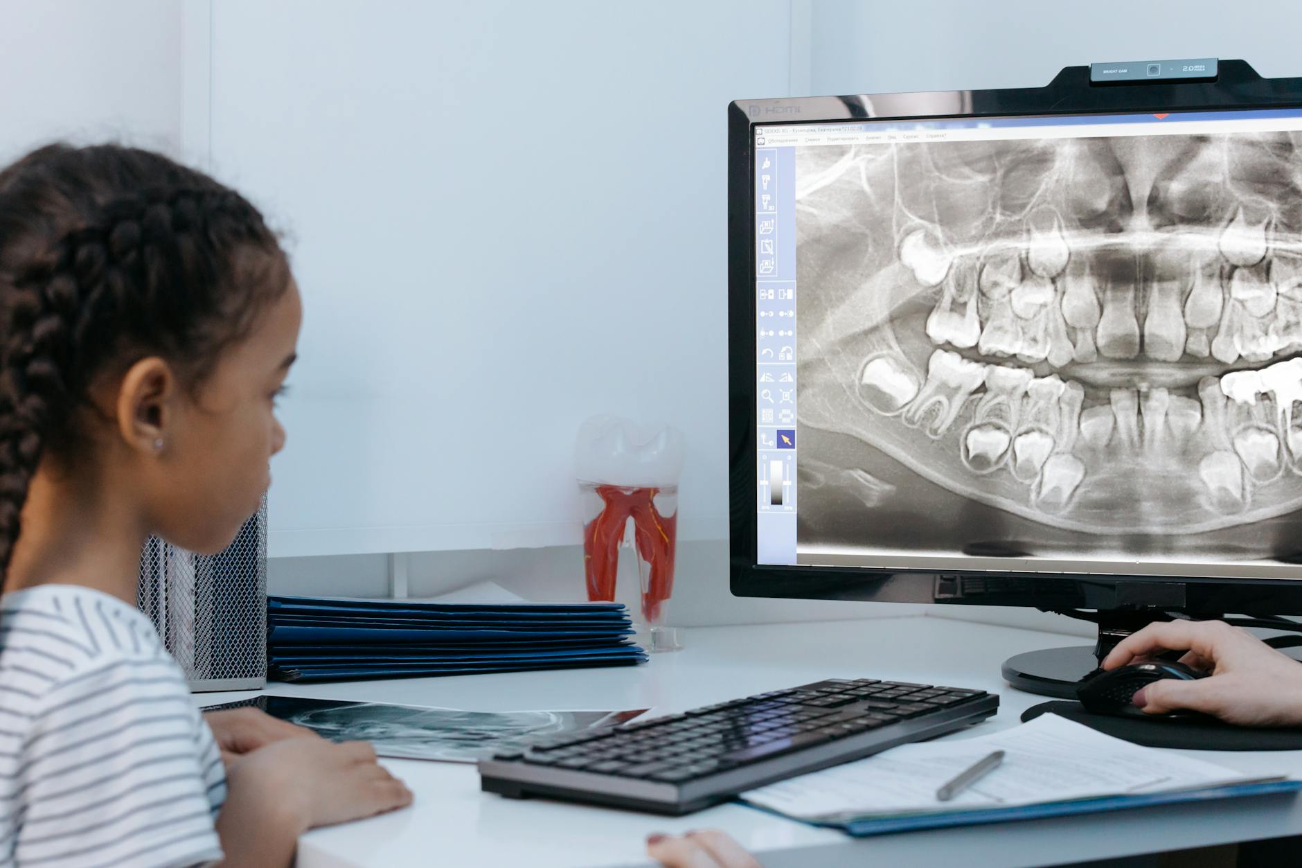
CT scans serve a different purpose. They excel at detecting erosions, bone sclerosis, and ankylosis—the fusing of joint surfaces—and they’re particularly useful for guiding interventional procedures when a doctor suspects advanced SI joint arthritis. Bone scans can identify stress fractures, inflammatory changes, infections, and tumors affecting the SI joint, though they’re reserved for situations where other imaging hasn’t clarified the diagnosis. The reality is that many imaging studies return normal results for both disc and SI joint problems, which is why imaging is a supporting tool rather than the final answer.
What Is the Gold Standard for Confirming SI Joint Pain?
When clinical examination and imaging leave the diagnosis uncertain, a diagnostic SI joint injection using X-ray fluoroscopy or ultrasound guidance represents the gold standard for confirming SI joint pain. This procedure involves injecting a small amount of local anesthetic directly into the SI joint and then observing whether the patient’s familiar pain diminishes. Pain relief is evaluated 20–30 minutes after the injection, with SI joint involvement confirmed if pain decreases by more than 75%.
The injection serves both diagnostic and therapeutic purposes. A positive response definitively establishes that the SI joint is responsible for the pain, which then guides treatment decisions moving forward. However, the procedure carries a small risk of infection or temporary increased pain, and some patients experience only partial relief even from a positive injection, suggesting that multiple pain generators might be contributing to their symptoms. For example, someone might receive an SI joint injection that relieves 80% of their pain but still experience residual discomfort from a minor disc bulge at L4-L5 that was incidentally noted on imaging but wasn’t causing significant symptoms until the SI joint pain resolved.
Why Do Standard MRI and X-Ray Often Appear Normal?
One of the most frustrating aspects of SI joint dysfunction is that standard MRI and X-ray imaging frequently appears completely normal despite the patient experiencing significant pain. This happens because mild to moderate SI joint inflammation, ligamentous instability, and early degenerative changes often don’t create enough anatomical disruption to show clearly on conventional imaging. A patient might undergo MRI, see a report saying “no abnormalities detected,” and leave the appointment feeling dismissed or confused. This is where the diagnostic injection becomes valuable—and why clinical examination tests remain so important.
A warning for patients: don’t assume that a normal MRI rules out SI joint dysfunction. The imaging is normal, but your pain is very real, and the diagnosis simply requires a different approach than looking at pictures. Some people pursue unnecessary spinal surgery for a disc herniation that appeared on imaging, only to discover after surgery that their SI joint dysfunction was the actual pain generator, and the disc herniation was incidental. This reinforces why doctors must integrate clinical findings with imaging rather than relying on imaging alone.

How Does Treatment Differ Based on the Diagnosis?
Once a diagnosis is confirmed, the treatment paths diverge significantly. Disc herniation treatment traditionally involves physical therapy, anti-inflammatory medications, and activity modification, with injection therapy or surgery reserved for cases where conservative care fails over several months. SI joint dysfunction treatment focuses on stabilization exercises, manual therapy to restore joint mechanics, and often SI joint belts or taping that provide external stabilization.
A practical example: two patients might both report lower back pain and appear to have similar symptoms initially. Patient A receives imaging showing a large disc herniation, begins a conservative program of core strengthening and anti-inflammatory medication, and gradually improves over eight weeks. Patient B receives an SI joint injection confirming SI joint dysfunction, starts a stabilization program with an SI belt, and within three weeks experiences marked improvement because the treatment directly addresses the joint’s instability. The diagnostic distinction allows doctors to prescribe targeted, effective treatment rather than generic back pain management.
What’s the Long-Term Outlook for Each Condition?
Disc herniation has a relatively well-understood natural history. Most people experience gradual improvement over three to six months with conservative care as the herniated material resorbs and inflammation subsides. However, recurrent herniation affects a percentage of patients, and some face chronic pain requiring long-term management strategies. SI joint dysfunction, when properly diagnosed and treated, often responds well to conservative care, though patients frequently require ongoing maintenance exercises to prevent recurrence.
The long-term outlook is generally favorable for both conditions when the diagnosis is accurate and treatment is appropriately matched to the problem. As our understanding of spine pain improves, diagnostic techniques continue to evolve. Emerging research into advanced imaging and more refined clinical testing protocols may eventually simplify the diagnostic process and reduce the number of patients who suffer through prolonged misdiagnosis. For now, the combination of careful clinical examination, selective imaging, and when necessary, diagnostic injection remains the most reliable method for distinguishing these two common causes of lower back pain.
Conclusion
Distinguishing disc pain from SI joint pain requires doctors to move beyond standard imaging and rely on a combination of specialized physical examination tests, targeted imaging, and diagnostic injections to reach a confident diagnosis. SI joint dysfunction accounts for a substantial portion of chronic lower back pain cases, yet its ability to masquerade as disc herniation on standard imaging makes accurate diagnosis crucial for effective treatment.
If you’re experiencing lower back pain that hasn’t improved with initial treatment or appears normal on standard imaging, ask your doctor about SI joint dysfunction as a potential diagnosis. Request physical examination testing and consider a diagnostic injection if initial tests suggest SI joint involvement. Understanding the true source of your pain is the essential first step toward targeted treatment and meaningful recovery.