The sacroiliac joint test most clinicians rely on is the FABER test (also called Patrick’s test), a simple physical examination where the patient lies on their back, one foot is placed on the opposite knee, and the examiner applies gentle pressure to the raised knee toward the floor. This position stresses the SI joint and sacroiliac ligaments; if a patient experiences pain in the lower back or buttock area during this maneuver, it suggests SI joint dysfunction or inflammation. For patients in dementia care settings, where mobility and fall risk assessment are critical parts of overall health management, understanding SI joint function matters because sacroiliac pain or instability can affect gait, balance, and quality of life—especially in older adults who are already at higher risk for falls and functional decline. This article covers what the SI joint is, why clinicians test it, how the FABER test works, its limitations, and what results might mean for patient care planning.
Table of Contents
- What Is the Sacroiliac Joint and Why Do Doctors Test It?
- How the FABER Test Is Performed and What It Measures
- Other SI Joint Tests Clinicians Use in the Clinic
- Interpreting FABER Test Results and What They Mean for Treatment
- Limitations of Clinical SI Joint Testing and When Imaging Is Needed
- SI Joint Testing in Older Adults and Dementia Care Contexts
- The Role of SI Joint Assessment in Comprehensive Mobility Evaluation
- Conclusion
What Is the Sacroiliac Joint and Why Do Doctors Test It?
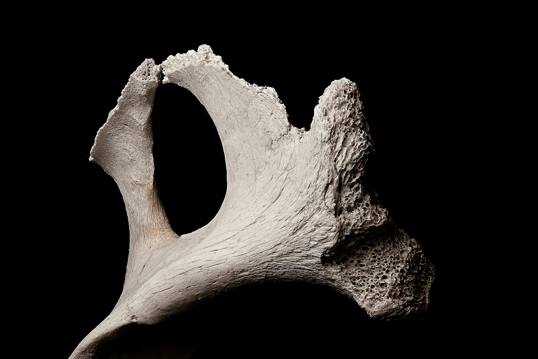
The sacroiliac joint is where the sacrum (the base of the spine) connects to the pelvis on each side. Unlike the spine, which has considerable mobility, the SI joints are designed to be relatively stable—they transfer force and weight between the upper body and legs during walking, standing, and sitting.
When these joints become inflamed, misaligned, or unstable, patients often experience sharp pain in the lower back, buttocks, or even down the thigh, and their movement patterns change. Doctors test the SI joint because SI joint dysfunction affects roughly 15-30% of people with chronic lower back pain, and identifying it changes treatment approach: exercises for SI joint stabilization look different from exercises for a herniated disc, for example. In elderly patients, particularly those with dementia who may have difficulty communicating pain or reporting symptoms accurately, clinical SI joint assessment helps clinicians understand why a patient’s mobility has declined or why they’re favoring one leg.
How the FABER Test Is Performed and What It Measures
The FABER test is performed with the patient supine (lying on their back). The examiner flexes the patient’s hip and knee on one side, then externally rotates the hip so the ankle rests on top of the opposite knee, creating a figure-four shape—this is why it’s sometimes called the figure-four test. From this position, the examiner applies gentle downward pressure on the raised knee.
The test checks whether movement and pressure in this position reproduce the patient’s symptoms, particularly pain felt over the SI joint region, buttock, or referred pain in the groin. A positive test doesn’t definitively diagnose SI joint dysfunction on its own; it’s most meaningful when paired with other clinical findings like palpation over the SI joint, patient history, and other provocative tests. However, if X then Y: if a patient has positive FABER test results combined with morning stiffness that improves with activity and pain that worsens with standing on one leg, the likelihood of SI joint involvement is much higher than FABER results alone would suggest.
Other SI Joint Tests Clinicians Use in the Clinic
While FABER is the most commonly used, clinicians often employ several other tests to triangulate a diagnosis. The SI compression test (also called the SI distraction test) involves applying direct compression or distraction force to the SI joints while the patient is sidelying or standing, which further stresses the joint capsule and ligaments.
Gaenslen’s test positions the patient near the edge of a table with one leg hanging off; the examiner applies downward pressure on the extended leg, creating a shearing stress across the SI joint. The NAAT (Numeric Analog Anterior-Posterior Test, though less commonly used than the others) involves measuring sacral motion with palpation. For dementia care patients who may struggle with positioning or cooperation, simpler tests like SI compression may be preferable to more complex maneuvers, and clinicians often rely more heavily on passive range-of-motion assessment and observation of how the patient moves than on a single definitive test.
Interpreting FABER Test Results and What They Mean for Treatment
A positive FABER test result means the patient experienced pain during the test, typically in the SI joint region or buttock, which suggests SI joint structures are irritated. However—and this is an important caveat—FABER has moderate sensitivity and specificity; some patients with true SI joint dysfunction may have a negative FABER, while others with pain in the hip or groin may have a positive test from hip joint involvement rather than SI joint pathology.
This is why skilled clinicians use FABER as one piece of information rather than a standalone diagnostic tool. When FABER is positive and other findings align (pain location, aggravating factors like stair climbing or rolling over in bed, palpation tenderness over SI joint markers), clinicians typically recommend SI joint-focused treatment: physical therapy for stabilization exercises, possible SI joint bracing, and activity modification. In dementia care settings, positive SI joint findings often mean adjusting mobility assistance techniques and physical therapy focus to prevent further deterioration and reduce pain.
Limitations of Clinical SI Joint Testing and When Imaging Is Needed
Clinical tests like FABER cannot determine whether SI joint pain is coming from the joint capsule, the ligaments, or referred pain from nearby structures like the piriformis muscle. This is a real limitation: two patients with identical positive FABER results may require completely different treatments depending on the actual anatomical source of pain.
Additionally, some patients (particularly those with severe osteoarthritis, pregnancy-related pelvic changes, or certain inflammatory conditions) may have positive SI joint tests that don’t fully explain their functional limitations. If conservative treatment (physical therapy, stabilization exercises) doesn’t improve symptoms over 4-6 weeks, or if neurological symptoms develop (numbness, weakness below the knee), imaging like X-rays or MRI may be needed to rule out other pathology. For dementia patients who have difficulty reporting precise pain location or progression, clinicians should watch for functional decline or gait changes that don’t respond to expected treatment as a signal that further investigation is warranted.

SI Joint Testing in Older Adults and Dementia Care Contexts
In older adults, SI joint dysfunction is often overlooked because lower back pain is attributed solely to degenerative disc disease or spinal stenosis, even when SI joint involvement is contributing. Testing SI joint function becomes especially valuable in dementia care because these patients may not communicate pain clearly, and clinicians must rely on observed movement patterns, expressions of discomfort, and family reports of functional changes.
An older person with dementia who suddenly becomes reluctant to stand, walks with an asymmetrical gait, or expresses pain when being transferred may be experiencing SI joint dysfunction—and recognizing this can direct physical therapy and daily care activities appropriately. One specific example: a patient with moderate dementia who was becoming increasingly sedentary was found to have a positive SI compression test and mild SI joint inflammation on imaging; once stabilization exercises and modified positioning during care were implemented, the patient’s mobility and willingness to participate in activities improved noticeably.
The Role of SI Joint Assessment in Comprehensive Mobility Evaluation
SI joint testing is increasingly recognized as a standard component of comprehensive lower-back and pelvic assessment, not just an afterthought or specialist concern. For dementia care teams, including SI joint screening in routine mobility assessments can identify a treatable source of pain and functional decline that might otherwise be missed or attributed to cognitive or behavioral issues.
As research continues to clarify the relationship between SI joint stability and overall spinal health, physical therapy approaches that address SI joint stabilization alongside spine mobility are becoming more evidence-based. In future care practices, particularly for aging populations with neurological conditions, SI joint assessment and stabilization may be integrated earlier into prevention programs to help maintain mobility and independence longer.
Conclusion
The FABER test (Patrick’s test) is the sacroiliac joint test most commonly used by clinicians because it’s simple to perform, requires no equipment, and effectively stresses the SI joint structures. While FABER alone cannot definitively diagnose SI joint dysfunction, it provides valuable clinical information when combined with patient history, pain location, and other physical findings. For dementia care settings where patients may have difficulty communicating symptoms and maintaining mobility is critical to quality of life, recognizing SI joint involvement can change the course of physical therapy, pain management, and daily care strategies.
If a patient experiences pain during SI joint testing or shows signs of SI joint dysfunction (pain with standing on one leg, asymmetrical gait, buttock pain), the next step is usually targeted physical therapy focused on stabilization exercises, pelvic bracing, and activity modification. If conservative care doesn’t improve symptoms within 4-6 weeks, or if symptoms worsen, imaging and further specialist evaluation may be warranted. Healthcare teams should maintain a low threshold for SI joint assessment in older adults and particularly in dementia care, where functional decline may be the primary signal of underlying pain or structural issues.