When a radiologist uses the word “encephalomalacia” in an MRI report, it means that an area of brain tissue has died and been replaced by cerebrospinal fluid — a permanent change that cannot be reversed. The term comes from the Greek for “brain softening,” and it describes the end result of liquefactive necrosis, a process in which dead neural tissue essentially liquefies and is gradually reabsorbed, leaving behind a fluid-filled cavity. It is not a diagnosis on its own; it is a descriptive finding that tells the reader something destructive has already happened to the brain at that location. To put this in concrete terms: if someone suffers a stroke that cuts off blood supply to a region of the left frontal lobe, the neurons in that area die within hours.
Over weeks and months, the dead tissue is cleared away, and the resulting space fills with cerebrospinal fluid. On a follow-up MRI, a radiologist would describe that cavity as encephalomalacia. The same outcome can follow traumatic brain injury, severe infection, brain surgery, or oxygen deprivation at birth. This article covers what encephalomalacia looks like on different MRI sequences, what causes it, what symptoms it can produce, and what options exist for managing its effects.
Table of Contents
- What Does Encephalomalacia Mean on a Brain MRI — and How Is It Identified?
- What Causes Encephalomalacia and Why Location Matters
- Symptoms Associated with Encephalomalacia
- Is Encephalomalacia Treatable — and What Can Be Done?
- Encephalomalacia vs. Other MRI Findings — Avoiding Confusion
- Encephalomalacia in the Context of Dementia and Aging
- What to Expect Going Forward After an Encephalomalacia Finding
- Conclusion
- Frequently Asked Questions
What Does Encephalomalacia Mean on a Brain MRI — and How Is It Identified?
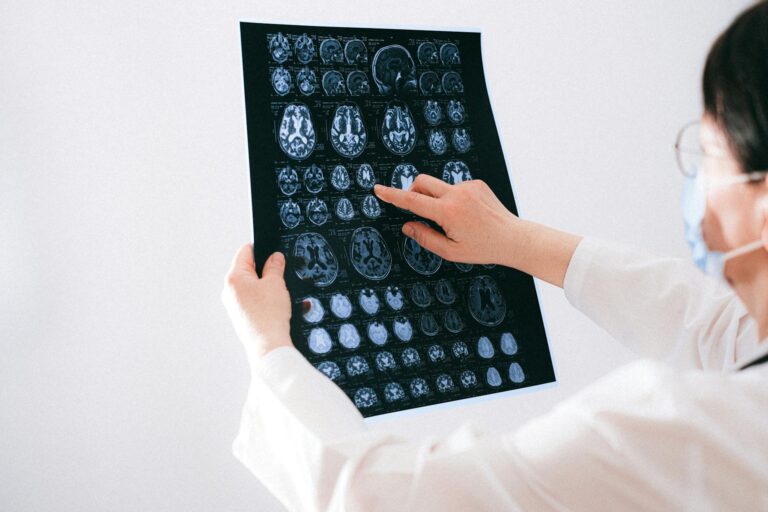
On an mri scan, encephalomalacia has a distinctive appearance that experienced radiologists recognize across multiple imaging sequences. On T1-weighted images, the affected area appears hypointense — meaning it shows up darker than surrounding healthy tissue. On T2-weighted images, it appears hyperintense, or brighter than normal. These two findings together strongly suggest the presence of cerebrospinal fluid where brain tissue once was, since CSF behaves in a predictable way on standard MRI sequences. The most diagnostically useful sequence is often T2 FLAIR, which stands for Fluid-Attenuated Inversion Recovery. This sequence is specifically designed to suppress the bright signal of free fluid, so cerebrospinal fluid appears dark.
If an area of the brain looks bright on T2 but dark on FLAIR, that strongly points to encephalomalacia rather than active inflammation or a new injury. The comparison matters clinically: surrounding gliosis — the scar tissue that forms around the damaged area — will appear as a high signal on FLAIR, creating a rim of brightness around the dark central cavity. This pattern helps radiologists distinguish old, stable tissue loss from an acute or subacute process. It is worth noting that the findings can appear focal, meaning confined to one specific region, or diffuse, meaning spread across wide areas of the brain. A person who had a small, localized stroke might show a single small focus of encephalomalacia near the internal capsule. A person who suffered severe traumatic brain injury or prolonged oxygen deprivation might show diffuse bilateral changes across multiple lobes.

What Causes Encephalomalacia and Why Location Matters
The most common cause of encephalomalacia is ischemic or hemorrhagic stroke. When blood supply is cut off from a region of the brain, neurons begin dying within minutes, and if the damage is extensive enough, the area undergoes liquefactive necrosis over the following weeks. Hemorrhagic strokes — where a blood vessel ruptures — can also leave behind encephalomalacia after the blood is reabsorbed and the tissue at the center of the bleed is destroyed. Traumatic brain injury is another leading cause, and it has a characteristic pattern. Post-traumatic encephalomalacia tends to appear in the anteroinferior frontal lobes and the temporal lobes — the regions most susceptible to the shearing and contusion forces that occur when the brain moves against the interior of the skull. A person who suffered a severe car accident decades ago might have an MRI today that shows old encephalomalacia in these locations with no other abnormality, a silent record of past trauma. Infections including encephalitis and meningitis can destroy brain tissue through a combination of direct viral or bacterial damage and inflammatory injury.
Brain tumors, surgical trauma, and perinatal hypoxic-ischemic injury — oxygen deprivation during birth — round out the major causes. However, location is not just a radiological detail. Where the encephalomalacia sits in the brain determines almost everything about its clinical consequences. Damage in the motor cortex will produce weakness or paralysis. Damage in the language areas — Broca’s or Wernicke’s — can produce aphasia. Damage in the temporal lobes may contribute to memory problems or seizures. Damage in the frontal lobes can alter personality and executive function in ways that are sometimes more distressing to families than physical deficits. Encephalomalacia in a clinically “silent” area of the brain — such as certain white matter regions — may produce no noticeable symptoms at all.
Symptoms Associated with Encephalomalacia
The clinical effects of encephalomalacia range from none whatsoever to severe and life-altering, depending entirely on where the tissue loss has occurred and how much tissue was destroyed. Some people walk around with documented encephalomalacia on imaging and have no idea it is there, because the affected region is not critical to daily function or because the brain has partially compensated through neuroplasticity over time. This is particularly common when the finding is small, focal, and located in white matter rather than primary cortex. At the other end of the spectrum, significant encephalomalacia — particularly when it affects motor, sensory, or language areas — can produce lasting deficits. Motor weakness or paralysis, loss of sensation on one side of the body, seizures, cognitive decline, and behavioral changes are all possible consequences.
Seizures are a particularly common complication because the surrounding gliotic tissue creates abnormal electrical activity at the margin of the old injury. Children who developed encephalomalacia from perinatal injury may show developmental delays, cerebral palsy, or epilepsy early in life. Behavioral changes deserve special mention, especially in the context of a dementia care audience. Frontal lobe encephalomalacia — whether from trauma, stroke, or surgery — can mimic or contribute to dementia-like presentations: apathy, poor judgment, impulsivity, and emotional dysregulation. When an elderly person’s cognitive changes are investigated with an MRI and encephalomalacia is found, it raises the question of whether vascular disease or old injury is contributing to what might otherwise be attributed solely to Alzheimer’s pathology.

Is Encephalomalacia Treatable — and What Can Be Done?
There is no treatment that can reverse encephalomalacia or regenerate the tissue that has been lost. Once brain cells have died and been replaced by a fluid cavity, current medicine has no mechanism to restore what was there. This is a hard truth for patients and families who receive this information in a radiology report, often without adequate explanation. The tissue is gone; the goal shifts entirely to managing whatever symptoms arise from the damage. Seizure management is often the most urgent clinical priority. Anti-seizure medications can reduce the frequency and severity of epileptic episodes that arise from the gliotic tissue surrounding the encephalomalacic cavity.
The tradeoff here is that anti-seizure medications carry their own cognitive side effects, particularly in older adults — so the prescribing clinician must weigh seizure risk against the cognitive burden of the medication. Physical therapy addresses motor deficits when encephalomalacia affects the motor cortex or corticospinal tracts. Speech therapy can help patients with aphasia or swallowing difficulties resulting from damage to relevant cortical regions. Cognitive therapy and rehabilitation may help patients compensate for executive function or memory deficits, even though the underlying tissue loss is permanent. The honest comparison to understand here is this: treating encephalomalacia is more like managing the aftermath of a scar than treating an active disease. The injury is fixed. What is variable is how much functional recovery is possible through compensation, adaptation, and rehabilitation — and that depends significantly on the age of the patient, the extent of the damage, and how much time has passed since the initial injury.
Encephalomalacia vs. Other MRI Findings — Avoiding Confusion
One of the most common points of confusion in reading brain MRI reports is distinguishing encephalomalacia from other white matter findings. Reports frequently mention white matter hyperintensities, leukoaraiosis, or periventricular changes — terms that describe different degrees of small vessel disease and chronic ischemic changes but do not necessarily indicate that tissue has been fully destroyed. Encephalomalacia is the end-stage finding: the tissue is gone, replaced by fluid. White matter hyperintensities on T2, by contrast, reflect injury that may range from mild ischemic change to early demyelination, and many of these lesions do not progress to full encephalomalacia. The distinction matters because it affects the clinical conversation. A report that says “scattered white matter hyperintensities consistent with small vessel disease” is describing a common, often age-related finding with uncertain functional significance.
A report that says “encephalomalacia in the left posterior frontal lobe consistent with prior infarct” is describing a permanent structural deficit that explains, or at least contributes to, any symptoms in the corresponding functional domain. Patients and families who receive reports using both kinds of language without explanation can easily conflate the two and either minimize a significant finding or become unnecessarily alarmed by a minor one. A warning worth emphasizing: encephalomalacia is a retrospective finding. The MRI is showing you what has already happened, not an active process. This is important because patients sometimes assume that finding encephalomalacia on a scan means they are currently having a stroke or that their brain is currently deteriorating. The radiologist is reading permanent structural evidence of past injury — not an acute event. However, if new symptoms have developed, imaging showing old encephalomalacia does not rule out a new concurrent problem, and clinical correlation remains essential.

Encephalomalacia in the Context of Dementia and Aging
In older adults being evaluated for cognitive decline, encephalomalacia often appears alongside other age-related and vascular changes on MRI. Its presence raises an important question about the relative contributions of different pathologies to the clinical picture. A patient presenting with memory loss might have both Alzheimer’s pathology and evidence of old cortical strokes with resulting encephalomalacia — a combination that tends to accelerate the clinical course compared to either pathology alone.
For families managing a loved one’s dementia care, understanding that a portion of the cognitive decline may be related to structural brain loss — rather than solely to protein accumulation — can inform care decisions. Vascular risk factor management (blood pressure, cholesterol, blood sugar) becomes even more important when existing encephalomalacia signals that the brain has already sustained vascular injury. While the lost tissue cannot be recovered, reducing the risk of further strokes is a meaningful and achievable goal.
What to Expect Going Forward After an Encephalomalacia Finding
The prognosis after encephalomalacia is largely determined by what happened before the scan, not after it. If the underlying cause — a stroke, an infection, a traumatic injury — has resolved and is unlikely to recur, then the encephalomalacia represents a stable endpoint. The fluid-filled cavity does not grow, spread, or evolve into something more dangerous on its own. Follow-up MRI may be recommended to confirm stability, but in many cases the finding is simply documented and monitored.
Where ongoing vigilance matters most is in cases where the cause of the original injury is not fully controlled. Someone whose stroke resulted from unmanaged atrial fibrillation or uncontrolled hypertension remains at risk for further events that could create additional areas of encephalomalacia. In those situations, the imaging finding should function as an urgent prompt to address the underlying vascular risk. The brain has limited tolerance for repeated insults, and each new area of tissue loss carries the potential for compounding functional decline.
Conclusion
Encephalomalacia on a brain MRI is a radiological description of permanent brain tissue loss — a region where neurons have died, the tissue has liquefied, and cerebrospinal fluid has taken its place. It is identified by characteristic signal changes across multiple MRI sequences, most definitively by a dark signal on T2 FLAIR surrounded by a bright rim of gliosis. The causes are varied — stroke is the most common, followed by traumatic brain injury, infection, and perinatal injury — but the outcome is always the same: irreversible structural change that medicine cannot undo.
What can be done is manage what follows. Seizure medications, physical therapy, speech therapy, and cognitive rehabilitation all target the functional consequences rather than the structural deficit itself. For patients and families navigating a report that contains this term, the most important initial step is a conversation with a neurologist who can interpret the finding in the context of symptoms, history, and overall brain health — not just the words on a radiology report. Understanding that the injury is old and stable, while remaining committed to preventing future vascular events, is the clearest path forward.
Frequently Asked Questions
Is encephalomalacia the same as a stroke?
No, but stroke is the most common cause of encephalomalacia. Encephalomalacia is the permanent residual finding — the cavity left behind after brain tissue has died and been reabsorbed. A stroke is the acute event; encephalomalacia is what the MRI shows months or years later.
Can encephalomalacia get worse over time?
The existing cavity is generally stable and does not expand on its own. However, the underlying cause — such as ongoing vascular disease — can produce new areas of injury that create additional encephalomalacia in other locations. Treating risk factors is essential to preventing new damage.
Does encephalomalacia always cause symptoms?
Not necessarily. Encephalomalacia in regions that are not critical to everyday function can be entirely asymptomatic. Whether symptoms occur depends on the location, size, and extent of the tissue loss, as well as how much the surrounding brain has been able to compensate.
Can the brain heal encephalomalacia?
No. Once brain tissue has undergone liquefactive necrosis and been replaced by cerebrospinal fluid, it cannot regenerate. Current medicine has no treatment that reverses the structural tissue loss. Management focuses entirely on symptom control and preventing further injury.
What does it mean if a child has encephalomalacia?
Encephalomalacia in children most often results from perinatal hypoxic-ischemic injury (oxygen deprivation around the time of birth), infections, or trauma. The consequences depend on the location and extent of the damage and may include developmental delays, cerebral palsy, or epilepsy.
Should I be worried if my MRI report mentions encephalomalacia?
The finding always warrants a conversation with a neurologist, but it does not necessarily indicate an active or worsening problem. In many cases it reflects an old, stable injury. Your doctor can explain what the finding means in the context of your specific symptoms and medical history.