Eye biomarkers are measurable changes in the eye that can signal early brain degeneration associated with dementia. Unlike invasive brain imaging or cognitive tests, these biomarkers can be detected through non-invasive eye scans—making them potentially powerful tools for identifying people at risk before memory loss becomes noticeable. Research has shown that the retina, the light-sensitive tissue at the back of the eye, shows structural changes in people with Alzheimer’s disease years before symptoms appear, offering a window into what’s happening inside the brain long before traditional diagnosis.
For dementia research, eye biomarkers solve a real problem: most current diagnostic methods either come too late (after significant cognitive decline) or require expensive, specialized brain imaging. An ophthalmologist can measure retinal thickness, detect changes in blood vessel patterns, or identify protein buildup in the eye during a routine examination. These observations may help researchers understand who will develop dementia, track disease progression, and eventually test whether new treatments slow brain decline.
Table of Contents
- Why Does the Eye Reflect What’s Happening in the Brain?
- What Eye Biomarkers Have Researchers Actually Discovered?
- How Are Researchers Measuring These Eye Biomarkers in Practice?
- Could Eye Biomarkers Become a Screening Tool for Dementia Risk?
- What Are the Key Limitations of Eye Biomarkers Today?
- Retinal Amyloid and Tau Imaging: A Newer Frontier
- What Does This Mean for People Concerned About Dementia Risk Today?
- Frequently Asked Questions
Why Does the Eye Reflect What’s Happening in the Brain?
The retina is essentially an extension of the central nervous system—it’s made of the same type of tissue as the brain itself, and it shares many of the same biological processes. When proteins like amyloid and tau accumulate in the brain during Alzheimer’s disease, similar accumulation occurs in the retina. This parallel process means the eye becomes a visible mirror of brain pathology. Researchers have found that people with cognitive decline or Alzheimer’s diagnosis show measurable thinning of the retinal nerve fiber layer compared to cognitively normal adults, a finding that has held up across multiple studies and imaging technologies.
The blood vessels in the retina also provide critical clues. In dementia-prone brains, the small blood vessels throughout the body—including those in the eye—undergo changes related to vascular damage and reduced blood flow. Optical coherence tomography angiography (OCTA), a specialized eye-imaging technique, can reveal these subtle vascular changes without injecting any dye or contrast agent. Some research suggests that retinal vessel narrowing and irregular blood flow patterns may precede memory loss by years, making them potential early warning signs before an individual seeks medical attention for cognitive concerns.
What Eye Biomarkers Have Researchers Actually Discovered?
The most consistent finding has been thinning of the retinal nerve fiber layer (RNFL) and the inner plexiform layer in people with Alzheimer’s disease. Multiple imaging studies using optical coherence tomography (OCT) have documented these structural changes, and the thickness appears to correlate with severity of cognitive impairment. One landmark study found that people with mild cognitive impairment—a transitional stage between normal aging and dementia—showed intermediate levels of retinal thinning, suggesting the changes progress alongside brain decline. However, there’s an important limitation: retinal thinning isn’t specific to Alzheimer’s disease.
Similar changes have been observed in other neurodegenerative conditions, including Parkinson’s disease and multiple sclerosis. This means an abnormal eye biomarker suggests neurodegeneration is occurring, but doesn’t pinpoint which condition. Additionally, not all people with Alzheimer’s pathology show retinal changes, and some people with retinal changes never develop dementia, which means eye biomarkers cannot yet serve as definitive standalone diagnostic tools. The findings are promising for research settings, but clinicians cannot yet use a retinal scan to tell an individual patient whether they will or will not develop dementia.
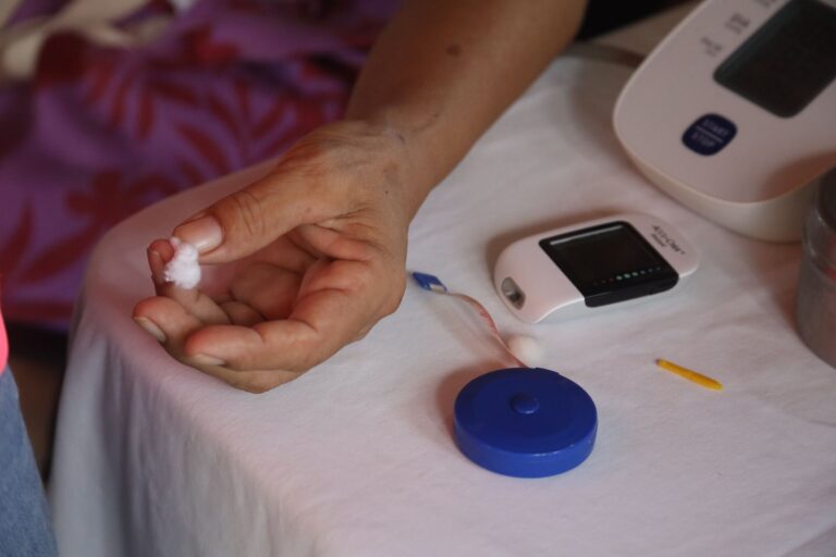
How Are Researchers Measuring These Eye Biomarkers in Practice?
Optical coherence tomography (OCT) has become the primary research tool for measuring retinal thickness and structure. The device works by bouncing light waves off the retina and creating a detailed cross-sectional map, much like an ultrasound but using light instead of sound. Patients sit in front of the machine, look at a fixed point, and the scan takes less than five minutes—far simpler than a brain MRI.
The resulting images show individual retinal layers in micrometers of detail, allowing researchers to measure changes that would be invisible to the human eye during a routine eye exam. Optical coherence tomography angiography (OCTA) extends this capability by visualizing the tiny blood vessels in the retina without requiring dye injection. This technique reveals blood flow patterns and vessel density, which researchers have found to be reduced in people with mild cognitive impairment and Alzheimer’s disease. In a study comparing people with Alzheimer’s to healthy controls, those with Alzheimer’s showed measurably lower retinal vessel density, a finding that could potentially help identify at-risk individuals in future screening programs.
Could Eye Biomarkers Become a Screening Tool for Dementia Risk?
The potential clinical application is compelling: if eye biomarkers could reliably predict who will develop dementia, a simple ophthalmology visit could identify high-risk individuals years before memory problems surface. This would allow people to start preventive interventions earlier—whether lifestyle changes, clinical trials, or future disease-modifying treatments. For healthcare systems, adding a retinal scan to routine eye exams would be far less expensive and invasive than requiring everyone to undergo PET imaging or spinal fluid testing to assess dementia risk.
But significant hurdles remain before this becomes standard practice. The relationship between eye biomarkers and actual dementia development has been documented in small research studies, but large population-based screening studies are still needed to establish how well these markers predict future disease in diverse age groups and ethnicities. The tradeoff is between early detection and false positives: if retinal changes are common in cognitively normal older adults who never develop dementia, then screening could create unnecessary anxiety. Researchers must determine whether biomarker changes are specific enough and prognostic enough to justify using them for identifying at-risk individuals in the general population.
What Are the Key Limitations of Eye Biomarkers Today?
One critical limitation is that eye biomarkers capture a snapshot of retinal structure and blood flow, but they don’t directly measure the biological hallmarks of dementia—amyloid and tau proteins. Researchers have inferred that retinal changes reflect underlying brain pathology based on studies in deceased patients and those undergoing PET imaging, but the eye scan itself isn’t directly visualizing the proteins driving neurodegeneration. Additionally, the same retinal changes occur in other conditions affecting the brain and blood vessels, so abnormal eye biomarkers lack specificity. A person with retinal nerve fiber layer thinning might have Alzheimer’s disease, vascular dementia, Parkinson’s disease, or simply advanced age-related changes—the eye biomarker alone cannot distinguish between these conditions.
Another warning: most current research has been conducted in specialized research centers with expensive OCT machines operated by trained technicians. Generalizing these findings to standard clinical practice in local ophthalmology offices is not straightforward. Different OCT machines can produce slightly different measurements, image quality varies based on patient factors like pupil size and eye clarity, and there is no agreed-upon diagnostic threshold—no universal “normal” versus “abnormal” retinal thickness that would apply to all people. Until these technical and standardization issues are resolved, eye biomarkers will remain primarily research tools rather than clinical screening instruments.
Retinal Amyloid and Tau Imaging: A Newer Frontier
More recent research has moved beyond measuring structural changes to actually visualizing amyloid and tau proteins in the retina using specialized fluorescent imaging techniques. Some companies have developed retinal imaging methods that can detect amyloid accumulation in the eye, potentially offering a direct biomarker of the same pathology occurring in the brain. If validated, this approach could solve the specificity problem by confirming the presence of Alzheimer’s-related proteins rather than just inferring neurodegeneration from structural changes.
However, retinal amyloid imaging is still in early research phases, and it’s unclear whether it will prove practical for widespread screening. The imaging technology is more complex than standard OCT, may require special eye drops or contrast agents, and will likely be more expensive. The evidence that retinal amyloid mirrors brain amyloid is still being established, and it remains unknown whether everyone with retinal amyloid will develop dementia or cognitive decline.
What Does This Mean for People Concerned About Dementia Risk Today?
For most individuals, getting eye biomarker screening through a routine eye exam is not yet possible or recommended. Ophthalmology practices don’t routinely perform the specialized OCT scans or OCTA imaging used in research, and even if they did, there is no established protocol for interpreting results or recommending interventions based on abnormal findings. People concerned about dementia risk should focus on modifiable factors with proven evidence: managing cardiovascular risk factors like blood pressure and cholesterol, staying cognitively and socially active, exercising regularly, maintaining a healthy diet, and getting adequate sleep.
The research on eye biomarkers is advancing, and someday these tests may become part of standard cognitive risk assessment. Several clinical trials are currently incorporating retinal imaging to track whether eye biomarker changes correlate with cognitive decline over time, which will help establish whether they can reliably predict future dementia. Until then, eye biomarkers remain a promising research avenue rather than a clinical tool, and people with family history of dementia or cognitive concerns should discuss standard cognitive and cardiovascular screening with their primary care doctor.
Frequently Asked Questions
Can I get my eye biomarkers screened at my eye doctor’s office right now?
Not yet. Specialized OCT and OCTA imaging for research are available only in research centers and some academic medical centers, not in routine eye care practices. Standard eye exams check eye health and vision, not dementia biomarkers.
If my eye scan shows retinal thinning, does that mean I will definitely develop dementia?
No. Retinal thinning can indicate several conditions and is not specific to dementia. Many people with retinal changes never develop cognitive decline. Eye biomarkers are useful for research but cannot predict individual outcomes at this time.
Are eye biomarkers more reliable than cognitive tests for detecting early dementia?
Not yet. Cognitive testing remains the standard method for identifying mild cognitive impairment and dementia. Eye biomarkers are complementary research tools that may eventually add to our ability to detect at-risk individuals, but they haven’t replaced current diagnostic methods.
If eye biomarkers predict dementia, can I start treatment to prevent it?
Even if someone is identified as high-risk based on eye biomarkers, there is no proven medication yet that halts Alzheimer’s disease before symptoms appear. Proven preventive strategies include cardiovascular health, cognitive engagement, exercise, diet, and sleep—which should be pursued regardless of biomarker status.
Why study the eye when dementia affects the brain?
The retina is part of the central nervous system and undergoes similar changes as the brain in dementia. A retinal scan is also safe, quick, non-invasive, and much cheaper than brain imaging, making it an attractive research tool.