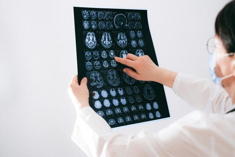
Periventricular white matter changes are areas of damage in the brain’s white matter — the tissue surrounding the ventricles, which are the fluid-filled spaces deep within the brain. On an MRI scan, these damaged areas appear as bright spots, which is why they are also called white matter hyperintensities (WMH) or, in older medical literature, leukoaraiosis. At the tissue level, these bright patches represent a mix of scarring, myelin degeneration, axonal loss, and tiny areas of infarction caused by chronically reduced blood flow. In plain terms, portions of the brain’s communication network have sustained injury, usually from years of inadequate circulation to small vessels.
The significance of these changes depends heavily on their extent. For a 70-year-old patient whose MRI shows a handful of small, punctate spots — the kind a radiologist might describe as “mild periventricular white matter changes” — this is often considered close to a normal finding for their age, with no immediate clinical consequences. For a patient whose MRI shows large, confluent regions of signal change spreading through the white matter, the picture is much more serious, carrying elevated risks of stroke, dementia, gait instability, and other neurological problems. This article explains what causes these changes, what the severity ratings actually mean, which symptoms they can produce, and what can realistically be done about them.
Table of Contents
- What Do Periventricular White Matter Changes Actually Mean on an MRI?
- What Causes Periventricular White Matter Changes?
- How Severity Is Classified and What Each Level Means
- What Symptoms Can Periventricular White Matter Changes Cause?
- The Relationship Between White Matter Changes and Dementia Risk
- Can Periventricular White Matter Changes Be Treated or Reversed?
- What Research and Future Developments May Change the Picture
- Conclusion
- Frequently Asked Questions
What Do Periventricular White Matter Changes Actually Mean on an MRI?
The ventricles are cavities inside the brain filled with cerebrospinal fluid. The tissue immediately surrounding them — the periventricular white matter — is dense with myelinated nerve fibers that carry signals between different brain regions. When blood supply to the small vessels feeding this area becomes chronically insufficient, the tissue degrades. Pathologically, what the radiologist sees as a bright spot on a T2-weighted or FLAIR MRI sequence corresponds to a combination of gliosis (scarring by support cells), loss of the myelin sheath protecting nerve fibers, actual axonal death, small-scale infarctions, and enlargement of the tiny spaces around blood vessels called perivascular spaces. It is worth understanding that “white matter changes” is a descriptive term, not a diagnosis. The MRI finding tells you something has happened to the tissue; it does not by itself explain why.
A neurologist interpreting the report needs to consider the patient’s age, medical history, and symptom profile before drawing clinical conclusions. A 55-year-old with uncontrolled hypertension and moderate white matter changes is in a very different situation from a 40-year-old with the same imaging finding and no vascular risk factors — the latter warrants investigation for less common causes such as multiple sclerosis or other demyelinating conditions. The terminology itself can cause confusion. Radiologists use several overlapping terms: periventricular hyperintensities, white matter hyperintensities, white matter lesions, leukoaraiosis, and white matter disease. These all refer to the same category of MRI finding and the underlying tissue injury. Some reports also distinguish between lesions immediately adjacent to the ventricles (periventricular) and those farther into the white matter (deep white matter lesions), as they may have slightly different significance, but in practice they often coexist and share the same general causes.

What Causes Periventricular White Matter Changes?
The overwhelming majority of periventricular white matter changes in adults over 50 are caused by cerebral small vessel disease — chronic microvascular ischemic injury driven by damage to the tiny arterioles that supply the deep white matter. The periventricular region is particularly vulnerable because it sits at the end of long, narrow arterial branches with limited collateral circulation. When those vessels narrow or stiffen due to years of high blood pressure, high blood sugar, smoking, or high cholesterol, the tissue they supply becomes chronically underperfused and begins to degenerate. Research also points to blood-brain barrier dysfunction as a contributing mechanism: when the barrier breaks down, toxic protein fragments and inflammatory molecules accumulate in the white matter, compounding the injury from reduced blood flow. Because the periventricular white matter is so sensitive to vascular insufficiency, hypertension is considered the single most important modifiable risk factor. Studies consistently show that people with long-standing, poorly controlled blood pressure carry a substantially higher burden of white matter changes than age-matched individuals with normal blood pressure.
Diabetes, smoking, and elevated LDL cholesterol each add independent risk. Age also plays a significant role — the prevalence and extent of white matter changes increases steadily after age 50 and is nearly universal in some form by age 80, even in otherwise healthy individuals. However, if white matter changes appear in a younger patient or the distribution is atypical, clinicians must consider other causes. Multiple sclerosis produces white matter lesions, though they tend to have a different distribution and shape than vascular WMH. Normal pressure hydrocephalus and idiopathic intracranial hypertension can also produce periventricular signal changes. Certain genetic conditions, including CADASIL (cerebral autosomal dominant arteriopathy with subcortical infarcts and leukoencephalopathy), cause severe white matter disease in young adults with minimal conventional vascular risk factors. The warning here is straightforward: age-inappropriate or rapidly progressive white matter changes should not be automatically attributed to the usual small vessel disease without broader investigation.
How Severity Is Classified and What Each Level Means
Radiologists and clinicians use grading scales to characterize how extensive periventricular white matter changes are. The Fazekas scale is among the most widely used: Grade 0 indicates no lesions, Grade 1 refers to punctate (small, dot-like) lesions, Grade 2 indicates early confluent lesions beginning to merge together, and Grade 3 describes large, confluent lesions that spread broadly through the periventricular white matter. The Scheltens scale and Age-Related White Matter Changes (ARWMC) scale offer similar stratification. These ratings help clinicians communicate severity and track progression over time. Mild, punctate periventricular lesions — a Fazekas Grade 1 finding — are generally considered near-normal in older adults. Most people with this degree of change have no symptoms attributable to the white matter and are not at dramatically elevated near-term risk.
Moderate lesions, corresponding roughly to Fazekas Grade 2, are associated with subtle cognitive and functional changes. Patients may notice mild slowing of processing speed, mild memory inefficiency, or slight difficulties with executive functions like planning and mental flexibility. They are not meeting criteria for dementia, but neuropsychological testing often reveals measurable differences compared to peers without such changes. Severe, confluent lesions at Fazekas Grade 3 carry meaningfully elevated clinical risks. Research has consistently linked this level of white matter change to significantly increased risk of stroke, dementia — including Alzheimer’s disease — gait and balance impairment, urinary incontinence, and neuropsychiatric symptoms such as depression and emotional lability. A 2025 study published in Frontiers in Neurology confirmed that periventricular hyperintensity severity independently predicts worse outcomes following ischemic stroke, meaning patients with higher white matter change burden before a stroke tend to have poorer recovery trajectories than those with fewer changes, even when the stroke itself is similarly sized.

What Symptoms Can Periventricular White Matter Changes Cause?
The clinical presentation of periventricular white matter changes varies considerably depending on severity and location. Mild changes are often entirely asymptomatic and discovered incidentally — for instance, when a patient undergoes brain imaging for headaches or after a minor accident, and the radiologist notes “scattered periventricular white matter changes consistent with small vessel disease” as an aside. Moderate to severe changes are more likely to produce noticeable effects, though these can be subtle enough that patients attribute them to normal aging rather than a specific brain condition. Cognitive effects tend to cluster in the domains of processing speed and executive function rather than the episodic memory loss stereotypically associated with Alzheimer’s disease. A person with significant periventricular white matter changes might find themselves slower to respond in conversation, more easily overwhelmed by complex tasks, struggling to multitask effectively, or requiring more effort to concentrate.
Gait disturbances are another important symptom — the periventricular white matter carries fibers involved in motor coordination, and when they are damaged, patients can develop a distinctive slow, shuffling, wide-based gait that increases fall risk. Urinary urgency and incontinence are also associated with severe white matter disease, as the white matter pathways involved in bladder control become disrupted. There is a meaningful tradeoff in how clinicians communicate white matter change findings to patients. On one hand, appropriate disclosure motivates patients to address vascular risk factors aggressively. On the other hand, there is evidence that patients who are told they have “brain changes” sometimes develop anxiety and health preoccupation that itself impairs function and quality of life. The most constructive framing emphasizes that the changes reflect past vascular stress on the brain, that many people with similar findings live well for years or decades without severe consequences, and that the most effective response is rigorous management of modifiable risk factors — not worry.
The Relationship Between White Matter Changes and Dementia Risk
One of the most common questions patients have after receiving an MRI report is whether periventricular white matter changes mean they will develop dementia. The honest answer is nuanced. White matter changes, particularly when moderate to severe, are associated with increased risk of dementia over time — but association is not certainty, and the risk is probabilistic rather than inevitable. Research published in PMC/NIH databases has documented that periventricular white matter hyperintensity burden correlates with dementia progression, and severe WMH can itself cause a vascular dementia syndrome as accumulated damage to white matter networks disrupts cognitive function at a clinical level. The relationship with Alzheimer’s disease is complex.
Cerebrovascular disease and Alzheimer’s pathology frequently coexist in the same brain, and white matter changes may lower the threshold at which Alzheimer’s pathology becomes clinically symptomatic. In other words, a brain with significant white matter disease may show cognitive symptoms at a lower amyloid plaque burden than a brain with intact white matter — the two insults are additive. This means that managing cerebrovascular risk factors is not only relevant for vascular dementia prevention but may also delay or moderate the clinical expression of Alzheimer’s disease in susceptible individuals. A critical warning: the presence of white matter changes on MRI should not be used as the sole basis for a dementia diagnosis. Many people with substantial white matter change burden remain cognitively intact or only mildly impaired for the rest of their lives, while others with relatively modest changes can decline significantly. Dementia diagnosis requires a clinical evaluation that incorporates cognitive testing, functional assessment, history, and often additional biomarkers — the MRI finding is one piece of a larger picture, not a stand-alone verdict.

Can Periventricular White Matter Changes Be Treated or Reversed?
No currently available treatment can reverse existing periventricular white matter changes. Once the tissue has undergone gliosis and axonal loss, those structural changes persist. This is an important reality for patients to understand, because it shifts the entire management focus from treating existing lesions to preventing further accumulation. The clinical value of identifying white matter changes lies in using them as a visible marker — a concrete MRI finding that motivates aggressive attention to the modifiable risk factors driving the ongoing injury.
Blood pressure control is the most evidence-supported intervention. Reducing and maintaining blood pressure within normal range has been associated with slower progression of white matter changes over time, though ongoing research continues to evaluate how much progression can be slowed and over what timeframes. Blood sugar management in diabetic patients, cholesterol reduction, and smoking cessation complete the core risk factor modification strategy. For many patients, seeing the MRI images and understanding that these bright spots represent real tissue damage is motivating in a way that abstract risk statistics are not. Clinicians at Mayo Clinic Proceedings have emphasized that white matter hyperintensities, precisely because they are visible and measurable, serve as a powerful clinical tool to encourage the consistent lifestyle and medication adherence that prevents further harm.
What Research and Future Developments May Change the Picture
The field of white matter research has advanced considerably in the past decade, and several lines of investigation hold promise for changing how periventricular white matter changes are managed in the future. Researchers are exploring whether interventions targeting blood-brain barrier integrity — rather than simply downstream vascular risk factors — could reduce the toxic protein accumulation that contributes to white matter injury. There is also growing interest in whether anti-inflammatory therapies might slow the gliotic scarring process after small vessel injury occurs.
Neuroimaging technology itself continues to improve. Advanced diffusion tensor imaging (DTI) can detect microstructural disruption in white matter that appears normal on conventional MRI, potentially identifying patients at risk before visible lesions develop. If reliable biomarkers for white matter vulnerability can be established, there is a realistic prospect of intervening earlier — before the damage becomes visible — rather than managing it after the fact. For now, the most effective strategy remains early identification of vascular risk factors and their sustained control, a mundane prescription that nonetheless carries significant preventive power over a lifetime.
Conclusion
Periventricular white matter changes are areas of tissue damage in the brain’s deep white matter, caused most commonly by chronic small vessel disease driven by hypertension, diabetes, and other vascular risk factors. On MRI they appear as bright spots surrounding the ventricles, and their clinical significance ranges from near-normal age-related findings at the mild end to substantially elevated risks of stroke, dementia, gait problems, and cognitive decline at the severe end. No treatment reverses existing lesions, but aggressive management of vascular risk factors — particularly blood pressure — is well-supported as a strategy for slowing further progression.
For anyone who has received an MRI report mentioning these changes, the most productive response is to treat it as actionable information rather than a frightening verdict. Work with a physician to understand the severity grading, address any modifiable risk factors with genuine seriousness, and if cognitive or gait symptoms are present, seek a comprehensive neurological evaluation. The changes on the MRI represent what has already happened; what comes next depends in large part on decisions made now.
Frequently Asked Questions
Are periventricular white matter changes the same as white matter disease?
They describe the same underlying process. “White matter disease” is a broader clinical term, while “periventricular white matter changes” or “white matter hyperintensities” are more specific descriptive terms used in MRI reports. All refer to areas of damaged white matter tissue resulting from chronic vascular injury.
Is it normal to have periventricular white matter changes at age 70?
Mild, punctate white matter changes are considered near-normal in adults over 60 to 70 and increase in prevalence with age. They are not entirely benign even at mild levels, but they do not automatically indicate disease or predict cognitive decline. Moderate to severe changes at any age warrant clinical attention.
Can periventricular white matter changes get worse?
Yes. White matter changes are generally progressive, particularly when underlying vascular risk factors remain uncontrolled. Follow-up MRI over years often shows expansion of existing lesions and development of new ones. Risk factor management is the primary tool for slowing this progression.
Do periventricular white matter changes cause headaches?
Headaches are not a well-established direct symptom of periventricular white matter changes. Many patients whose MRI incidentally reveals these changes initially underwent imaging for headaches, but the headaches are typically attributable to other causes. If headaches are severe or progressive, a physician should evaluate the full clinical picture rather than attributing them solely to the white matter finding.
Should I be worried if my MRI says mild periventricular white matter changes?
Mild changes in an older adult are common and often require no specific treatment beyond regular vascular risk factor monitoring. They do serve as a prompt to ensure blood pressure, blood sugar, and cholesterol are well managed. A conversation with your physician about the finding in the context of your full health history is the appropriate next step.
Can young people get periventricular white matter changes?
Yes, though it is uncommon in younger adults without specific risk factors. When white matter changes appear in someone under 45, clinicians look more carefully for less common causes including multiple sclerosis, genetic conditions like CADASIL, migraine-related white matter changes, or other autoimmune and inflammatory disorders, rather than assuming typical small vessel disease.