Reviewed by the Help Dementia Editorial Team — our editors review every article for accuracy against guidance from the National Institute on Aging, the Alzheimer’s Association, and peer-reviewed sources.
Research into brain visualization has advanced dramatically over the past two years, fundamentally changing how scientists and clinicians can observe the human brain’s structure and function. Recent breakthroughs in imaging technology—from ultra-high-resolution MRI scanners to electron microscopy mapping and AI-powered visualization tools—now allow researchers to see brain tissue in unprecedented detail. For example, in May 2024, researchers using the NIH BRAIN Initiative created the highest-resolution map of human brain tissue ever produced, using electron microscopy to map a cubic millimeter of brain tissue at the subcellular level, revealing complexity previously impossible to visualize with conventional techniques.
These advances matter particularly for dementia research and early detection. When we can see the brain’s architecture and function more clearly, we gain insights into how diseases develop and progress at a cellular level. The improvements in visualization technology translate directly into better diagnostic tools and the potential for earlier intervention in conditions like Alzheimer’s disease and other forms of dementia.
Table of Contents
- How Can Advanced MRI Technology Reveal Microscopic Brain Details?
- What Are the Latest Breakthroughs in Brain Mapping Technology?
- How Do Researchers Now Capture Rapid Brain Cell Communication?
- Can Brain Imaging Now Diagnose Disease More Quickly?
- What Are the Limitations of Current Brain Visualization Technology?
- How Are Brain-Computer Interfaces Revealing Neural Function?
- What Does the Future of Brain Visualization Look Like?
- Conclusion
How Can Advanced MRI Technology Reveal Microscopic Brain Details?
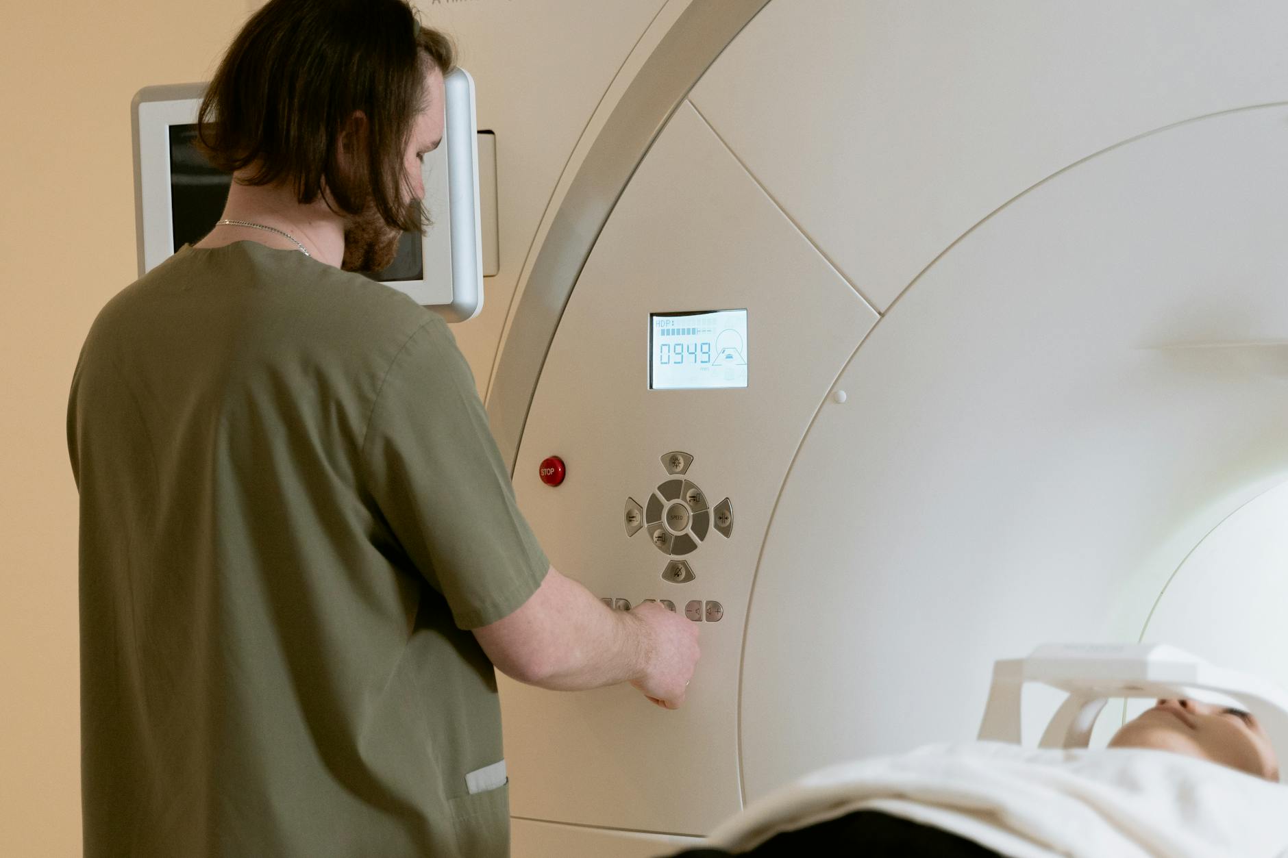
Recent breakthroughs in MRI technology have shattered the resolution limits that researchers have worked within for years. The newest generation of 7 Tesla MRI scanners records 10 times more detail than current 7T systems and over 50 times more detail than the standard 3T scanners found in most hospitals. These ultra-high-resolution scanners can now reveal functional MRI features as small as 0.4 millimeters—compared to the typical 2 to 3 millimeters visible on standard 3T fMRI scans.
What makes this advancement particularly significant is the increased signal-to-noise ratio, meaning researchers can distinguish actual brain activity from background interference. The NIH has specifically developed an ultra-high-resolution system called the Connectome 2.0 human MRI scanner, designed to reconstruct microscopic brain structures and identify the tiny connections that make up the brain’s “connectome”—the complete map of neural connections. However, this increased resolution comes with a tradeoff: scanning times remain lengthy, and the technology is expensive, limiting its availability primarily to research institutions.
What Are the Latest Breakthroughs in Brain Mapping Technology?
Artificial intelligence combined with traditional imaging has created new possibilities for analyzing brain scans. The NextBrain AI Atlas represents a major leap forward, enabling researchers to visualize the human brain in unprecedented detail down to hundreds of tiny subregions that were previously invisible on standard MRI scans. Rather than relying only on what human researchers could manually identify, AI systems now identify and map brain regions automatically, working at a pace and precision humans cannot match. Beyond MRI, researchers have developed alternative imaging approaches.
Scientists from USC and Caltech demonstrated functional photoacoustic computerized tomography (fPACT), which produced the first-ever images of human brain function created using this method. This technology offers highly detailed brain images without some of the traditional limitations of MRI, such as the need for magnetic fields and the time required for scanning. At the same time, the field continues to expand with new fluorescent nanobodies technology published in Nature Methods on April 22, 2026, which reduces background noise by up to 100-fold. This noise reduction enables much sharper visualization of where specific proteins are located in brain tissue and how they move and interact over time.
How Do Researchers Now Capture Rapid Brain Cell Communication?
Understanding how brain cells communicate happens at speeds that traditional imaging simply cannot follow. researchers at Johns Hopkins Medicine developed a “zap-and-freeze” method that solves this problem by using electrical stimulation to halt rapid communication between brain cells long enough to observe it. The technique allows researchers to capture interactions in living tissue from both mice and humans—activities that normally occur too quickly to track visually. This breakthrough opens doors to studying synaptic dysfunction in conditions where cellular communication breaks down, including in dementia-related diseases.
The implications for understanding neurological disease are substantial. Many age-related cognitive conditions, including Alzheimer’s disease, involve progressive breakdown in how neurons communicate. By capturing these interactions at the moment they occur, researchers can better understand what goes wrong in disease states and potentially identify intervention points. The challenge, however, is that this technique is still primarily a research tool, and scaling it for clinical diagnostic use remains years away.

Can Brain Imaging Now Diagnose Disease More Quickly?
Some of the most practical applications of enhanced brain visualization are already appearing in clinical settings. Multiple sclerosis, for instance, can now be accurately diagnosed using a specialized 8-minute MRI scan that employs T2*-weighted imaging to detect characteristic brain lesions that are centered on veins—a distinctive pattern of MS-related damage. This represents a significant advance for patients, as faster diagnosis means faster treatment initiation and better long-term outcomes.
The speed advantage cannot be overstated for clinical practice. Where a traditional diagnostic MRI workup might require 30 minutes to an hour of scanning time plus hours of radiologist review, newer focused protocols can deliver diagnostic results in minutes. For patients experiencing concerning neurological symptoms and for their families waiting for answers, this acceleration matters. However, the technology is not yet universally available, and access depends on whether imaging centers have been upgraded with the latest scanner hardware and protocols.
What Are the Limitations of Current Brain Visualization Technology?
While the advances in brain imaging are remarkable, significant challenges remain. Processing and interpreting the massive amounts of data generated by high-resolution imaging requires substantial computing resources and specialized expertise. When researchers map a cubic millimeter of brain tissue at the subcellular level, they generate petabytes of data—far more information than human researchers can analyze without AI assistance. This creates a bottleneck: the technology can now see more than we can easily understand.
Another limitation is that enhanced visualization does not automatically translate to clinical use. Many of the newest imaging breakthroughs are housed in specialized research centers and remain expensive and time-consuming. The ultra-high-resolution scanners require lengthy scan times and specialized protocols, making them impractical for widespread diagnostic screening. Additionally, visualizing structure does not always explain function—seeing where neurons are located is valuable, but understanding why they are communicating incorrectly in disease states requires additional research. For dementia research specifically, this means that even with perfect visualization of brain changes, translating those observations into treatments remains a separate, lengthy challenge.

How Are Brain-Computer Interfaces Revealing Neural Function?
Brain-computer interfaces have advanced alongside visualization technology, and the two technologies inform each other. A high-density brain-computer interface trial successfully decoded speech intentions at approximately 32 words per minute with remarkable accuracy, reading neural activity from a small implant and converting it into synthesized speech.
This breakthrough demonstrates not just the precision of neural recording but also what detailed brain visualization reveals: the specific neural pathways and firing patterns associated with speech and intention. For people with conditions that affect communication—including some forms of dementia or neurological disease—this technology hints at future possibilities. Understanding exactly which neurons encode speech and how they communicate allows researchers to develop interfaces that work with, rather than against, the brain’s natural organization.
What Does the Future of Brain Visualization Look Like?
Research teams at MIT have developed technology pipelines enabling high-resolution, high-throughput imaging of full hemispheres of human brains from both Alzheimer’s disease cases and control subjects. This whole-brain approach represents a shift from studying isolated regions toward comprehensive mapping.
As these capabilities mature, researchers will be able to compare brain structure and organization across hundreds of individuals, identifying subtle patterns that distinguish healthy aging from disease. The convergence of improved imaging, AI-powered analysis, brain-computer interfaces, and large-scale data collection is creating unprecedented opportunities for understanding neurological disease. Within the next several years, these research advances are likely to translate into improved screening tools, earlier diagnosis, and ultimately, better treatment strategies for conditions like dementia that currently have limited therapeutic options.
Conclusion
Research into brain visualization has entered a new era. From electron microscopy maps revealing subcellular detail to 7 Tesla MRI scanners capturing structures at 0.4-millimeter resolution, from AI atlases identifying previously invisible brain regions to “zap-and-freeze” methods capturing rapid cellular communication, the tools available to neuroscientists have expanded dramatically.
These advances represent not just incremental improvements but fundamental shifts in what can be observed and measured in the living and preserved human brain. For patients and families affected by dementia and other neurological conditions, these research breakthroughs eventually translate into better diagnosis, earlier detection, and the scientific foundation necessary for developing effective treatments. While many of these technologies remain in research settings today, the pace of translation from laboratory to clinic suggests that practical applications will continue to emerge in the coming years.