MRI scans detect herniated discs by using powerful magnetic fields and radio waves to create detailed cross-sectional images of your spine, allowing radiologists to visualize disc material that has shifted out of its normal position. Unlike X-rays, which only show bone, MRI captures soft tissue detail with such clarity that doctors can see the exact size and location of a disc bulge, whether it’s pressing on a nerve, and how much inflammation surrounds the damaged disc. A typical MRI of the lumbar spine will show 5-6 vertebrae stacked with their cushioning discs between them; when a disc herniates, you see the disc material protruding beyond its normal boundaries—sometimes appearing as a small bulge, sometimes as a fragment that has broken loose inside the spinal canal. This article explains how MRI technology detects herniated discs, why it’s considered the gold standard for diagnosis, how it compares to other imaging methods, what limitations doctors work around, and what your scan results actually mean for your treatment options.
Table of Contents
- How MRI Technology Visualizes a Herniated Disc
- MRI’s Advantage Over Other Imaging Methods for Herniated Discs
- Understanding What the MRI Report Actually Describes
- How MRI Compares to Physical Exams and Imaging Alternatives
- Limitations and Pitfalls in MRI Detection of Herniated Discs
- What to Expect During an MRI Scan for Spinal Imaging
- The Role of MRI in Modern Spine Care and Future Imaging
- Conclusion
How MRI Technology Visualizes a Herniated Disc
An MRI machine generates a strong magnetic field—typically 1.5 or 3 Tesla, measured in units of magnetic strength—that aligns hydrogen atoms in your body’s tissues. Radio waves then pulse through your spine, knocking these atoms out of alignment; when the radio waves stop, the atoms realign and emit signals that the machine detects and converts into images. Because disc material, nerve tissue, bone, and fluid all have different water and fat content, they emit slightly different signals, creating contrast that makes a herniated disc visible as a distinct structure pushing outward from its normal disc space.
The MRI captures images in multiple planes—axial (looking down from above), sagittal (looking from the side), and coronal (looking from front to back)—which gives your doctor a three-dimensional understanding of the herniation. A small posterolateral bulge at the L4-L5 level, for example, might appear as a subtle outpouching on a sagittal view but clearly visible on an axial slice where you can see exactly which direction it’s pushing and whether it contacts the nerve root. The detail is so precise that radiologists can measure the herniation in millimeters and determine whether the disc material is contained (still covered by the outer disc layer) or extruded (broken through that outer layer).

MRI’s Advantage Over Other Imaging Methods for Herniated Discs
Unlike X-rays, which show only bone and would miss a soft-tissue disc herniation entirely, MRI captures the disc and surrounding nerves and spinal cord with exceptional clarity. CT scans can show discs well but deliver more radiation and provide less soft-tissue contrast than MRI; they’re often reserved for patients who can’t have an MRI due to metal implants like pacemakers or metallic joint replacements. However, one critical limitation: MRI shows structural abnormality—the disc is herniated—but it cannot directly show whether that herniation is causing your pain.
A study in *Radiology* found that 30% of people without any back pain had incidental disc herniations on MRI, which means seeing a herniation doesn’t automatically mean it’s responsible for your symptoms. This distinction matters enormously in clinical practice. Two patients might have identical-looking herniations on MRI, but one experiences severe pain and the other feels nothing, because the herniation’s effect depends on its exact relationship to the nerve root—millimeter-level positioning and the individual’s anatomy. This is why your doctor also performs a physical exam, asks about your symptoms, and may order other tests like electromyography (EMG) if needed.
Understanding What the MRI Report Actually Describes
When you receive your MRI results, the radiologist’s report will describe the herniation using specific terminology that affects how seriously it’s taken. A “bulge” means the disc’s outer boundary extends evenly all around but the material hasn’t actually ruptured—less concerning and often self-resolving. A “protrusion” means the disc material has pushed outward but is still contained by the outer ligament.
An “extrusion” or “sequestration” means the disc material has broken through the outer layer and may migrate up or down the spinal canal, which is more likely to cause nerve irritation and warrant more aggressive treatment. The report will also specify the disc level—L5-S1 at the base of your spine, L4-L5 in the lower back, and so on—and whether it’s central, lateral, or posterolateral, which tells your doctor which nerves are at risk. A large left-sided L4-L5 herniation might compress the left L5 nerve root, causing pain, numbness, or weakness down the left leg; the same herniation on the right wouldn’t affect the left leg. This anatomical precision is one reason MRI changed spine care so dramatically when it became widely available in the 1980s—before that, doctors could only guess where a disc herniation was based on your symptoms.
How MRI Compares to Physical Exams and Imaging Alternatives
A skilled physical therapist or physician can often predict roughly where your herniation is by testing your reflexes, strength, and sensation—the straight-leg raise test, for example, often triggers pain if you have a lower lumbar herniation pressing on a nerve—but only MRI shows the actual structural damage. Some doctors start with imaging alternatives: ultrasound can show larger herniations but lacks detail; a CT scan takes less time than MRI and works for metal-implant patients but delivers radiation. However, if your symptoms don’t clearly point to a single disc level, or if you’re being considered for surgery, MRI is the standard because it shows the exact anatomy your surgeon needs to plan an operation.
One practical tradeoff: MRI is expensive (often $1,500–$4,000 out-of-pocket), time-consuming (30–45 minutes in a loud machine), and can trigger claustrophobia in some people. Many insurers now require doctors to try conservative treatment—rest, physical therapy, anti-inflammatory medication—before approving an MRI, which delays diagnosis but reduces unnecessary imaging. For uncomplicated back pain, this approach makes sense; for severe neurological symptoms like progressive weakness or loss of bladder control, an urgent MRI is justified because those signs suggest cord compression requiring prompt surgery.
Limitations and Pitfalls in MRI Detection of Herniated Discs
MRI is excellent at showing disc herniations but poor at assessing whether a disc is truly painful. Additionally, some herniations hide behind bone spurs or occur in regions of severe degenerative disc disease where the contrast between normal and herniated material becomes less obvious. If you have extensive arthritis or spinal fusion hardware from prior surgery, artifact—image distortion—can obscure the view.
In these cases, advanced imaging like high-resolution CT or even a surgical exploration might be needed for clarity. Another limitation: an MRI is a snapshot. A disc herniation can reabsorb over weeks or months without surgery, especially if the extruded fragment is loose in the spinal canal; your radiologist sees the herniation at the moment of imaging but can’t predict whether it will improve on its own or persist. This is why serial imaging—repeating the MRI after 6-8 weeks of conservative care—sometimes shows a smaller or resolved herniation, which changes the treatment plan dramatically from surgery to continued therapy.

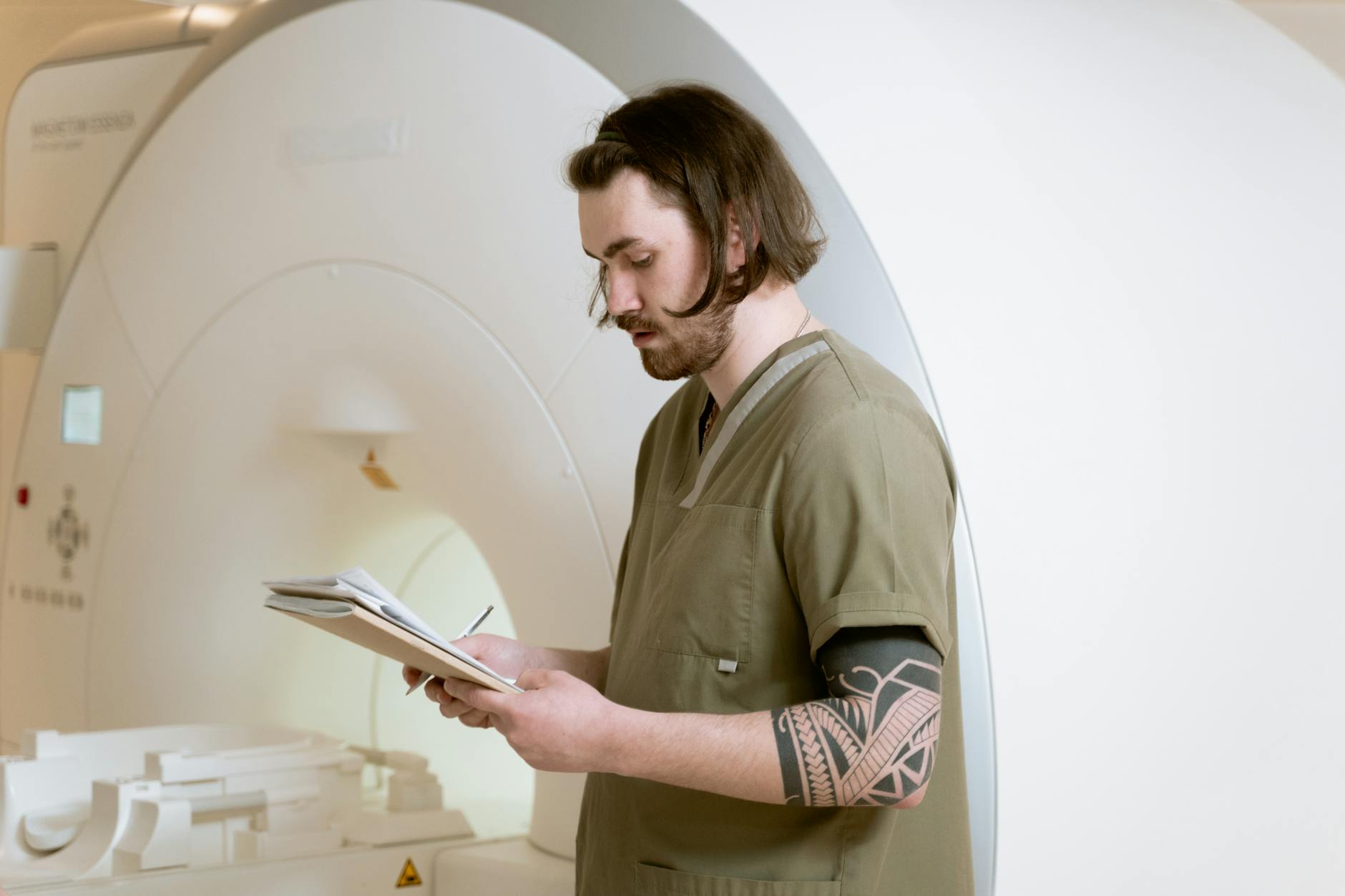
What to Expect During an MRI Scan for Spinal Imaging
When you arrive for a lumbar spine MRI, you’ll change into a gown and lie on a table that slides into the MRI bore, a cylindrical tube that can feel tight or claustrophobic for some people. The machine is loud—magnetic pulses create repetitive banging and buzzing sounds—and you’ll wear headphones and may hear the technician communicating. The scan takes 20–45 minutes depending on how many images are needed; you must remain perfectly still because any motion blurs the images.
Some facilities offer open MRI machines for claustrophobic patients, though they produce lower image quality; if you know you’re claustrophobic, mention it beforehand so the facility can schedule accordingly. Once the images are captured, a radiologist reviews them and generates a report—usually available within 24–48 hours—describing any herniations, their size, location, and relationship to nerves. Your primary care doctor or spine specialist will review the report and images with you, explaining what you do and don’t need to worry about and discussing treatment options.
The Role of MRI in Modern Spine Care and Future Imaging
MRI has been the standard for diagnosing herniated discs for over 30 years, but newer techniques are emerging. Diffusion tensor imaging (DTI) can assess nerve fiber integrity in cases of chronic compression, helping predict who will recover well with conservative care. Some research centers use functional MRI to correlate disc herniations with actual pain processing in the brain, though this remains experimental.
As MRI technology improves—7-Tesla machines offer higher resolution than standard 3-Tesla scanners—the ability to detect smaller herniations and earlier degenerative changes will increase, potentially shifting care toward earlier intervention in some cases. For now, if you’re experiencing back pain with nerve symptoms, an MRI remains the most reliable way to determine whether a herniated disc is the culprit and, if it is, exactly what your surgeon or therapist is dealing with. Advances in artificial intelligence are beginning to assist radiologists in analyzing MRI images faster and flagging subtle findings, which may make diagnosis quicker and more consistent in the coming years.
Conclusion
MRI scans detect herniated discs with exceptional clarity by creating detailed images of your spine’s soft tissues, showing the exact location, size, and direction of a disc bulge or extrusion. The scan provides the anatomical roadmap that doctors need to plan treatment, distinguish a real problem from an incidental finding, and explain your symptoms. However, seeing a herniation on MRI doesn’t automatically mean surgery is needed—many herniations improve with rest, physical therapy, and anti-inflammatory treatment.
If you’re scheduled for a spine MRI or recently received results showing a herniation, discuss the findings with your doctor to understand what the report means for your specific symptoms and treatment options. Conservative management works for the majority of herniated disc cases, and surgery is reserved for situations where neurological symptoms are severe or progressively worsening. MRI gives you and your doctor the information needed to make that decision confidently.