MRI can reveal brain changes commonly associated with Alzheimer’s disease, but it cannot confirm an Alzheimer’s diagnosis on its own. An MRI scan shows structural changes like shrinkage in certain brain regions, particularly the hippocampus, which plays a crucial role in memory formation. When a 68-year-old patient with progressive memory loss undergoes an MRI and doctors observe hippocampal atrophy along with other brain changes, that imaging provides important clues—but those clues must be interpreted alongside cognitive testing, medical history, and other diagnostic tools to reach an actual diagnosis.
The key distinction is that MRI shows what is happening to the brain’s structure, but it cannot show the underlying pathology that defines Alzheimer’s disease: the accumulation of amyloid-beta plaques and tau tangles. These protein deposits are the hallmark of the disease at the microscopic level and cannot be seen on standard MRI scans. This is why MRI functions as one piece of a larger diagnostic puzzle rather than a definitive test.
Table of Contents
- What Brain Changes Can MRI Detect in Alzheimer’s Disease?
- Which MRI Findings Are Most Specific to Alzheimer’s Disease?
- How Is MRI Integrated Into the Alzheimer’s Diagnostic Process?
- When Is an MRI Ordered for Suspected Alzheimer’s Disease?
- What Are the Key Limitations of MRI in Alzheimer’s Diagnosis?
- Can MRI Distinguish Alzheimer’s Disease From Other Types of Dementia?
- What New MRI Techniques Are Emerging for Better Alzheimer’s Detection?
- Frequently Asked Questions
What Brain Changes Can MRI Detect in Alzheimer’s Disease?
MRI is particularly useful for measuring brain atrophy—the shrinkage and loss of brain tissue that occurs in Alzheimer’s. The technology creates detailed images of brain structure by using magnetic fields and radio waves, allowing doctors to see if specific areas have become smaller than expected for a person’s age. One of the earliest and most consistent findings in Alzheimer’s disease is atrophy of the hippocampus, a seahorse-shaped structure deep within the brain that stores and retrieves memories. Research shows that people with Alzheimer’s often have a hippocampus that is 10 to 15 percent smaller than what’s typical for their age group.
Beyond the hippocampus, MRI can reveal widespread cortical atrophy—thinning of the brain’s outer layer—and enlargement of the ventricles, the fluid-filled spaces inside the brain. These changes occur because neurons die and are not replaced. Comparing an MRI from a patient with Alzheimer’s to one from a healthy person of the same age shows a striking difference: the Alzheimer’s brain has more visible gaps and spaces where tissue used to be. However, not everyone who has mild cognitive changes shows obvious atrophy on MRI, and some cognitively healthy older adults have atrophy that rivals that of people with cognitive decline, illustrating that brain shrinkage alone does not equal disease.
Which MRI Findings Are Most Specific to Alzheimer’s Disease?
Hippocampal atrophy is the MRI finding most closely linked to Alzheimer’s disease, but it is not unique to it. The same pattern can appear in other conditions, including vascular dementia, depression, normal aging, and even chronic stress. This overlap is a significant limitation: a doctor cannot simply see hippocampal shrinkage and declare that a patient has Alzheimer’s.
Other dementias and neurological conditions produce similar MRI patterns, making it essential to look at the whole clinical picture. researchers have identified patterns that increase the likelihood of Alzheimer’s, such as relatively preserved overall brain volume paired with selective shrinkage in the medial temporal lobe (the region containing the hippocampus), plus changes in specific cortical areas. When someone has these specific atrophy patterns plus cognitive test results pointing to memory loss and a family history of dementia, the probability of Alzheimer’s disease rises considerably. Yet even with these patterns, MRI cannot rule out other causes of dementia or distinguish Alzheimer’s from frontotemporal dementia, Lewy body dementia, or mixed pathology—situations where a patient actually has multiple types of brain disease at once.
How Is MRI Integrated Into the Alzheimer’s Diagnostic Process?
A doctor typically orders an MRI when someone presents with cognitive concerns to rule out other brain conditions that might be causing symptoms. A 72-year-old who comes in with memory problems needs an MRI partly to exclude treatable causes like a brain tumor, subdural hematoma (bleeding), normal pressure hydrocephalus (a condition where fluid buildup impairs cognition), or stroke. The MRI rules out these medical emergencies and structural problems that might be confused with Alzheimer’s.
Once other causes have been excluded, the MRI findings are considered alongside a neuropsychological test battery—formal cognitive testing that measures memory, language, attention, and reasoning—plus information from family members about how the person’s thinking has changed over time. Blood tests for biomarkers like phosphorylated tau and amyloid levels have become increasingly important in recent years and often take precedence over MRI in modern diagnostic criteria. The doctor weighs the atrophy pattern, the cognitive profile, the biomarker results, and the clinical history to build a diagnostic picture. This multimodal approach is far more reliable than any single test, including MRI alone.
When Is an MRI Ordered for Suspected Alzheimer’s Disease?
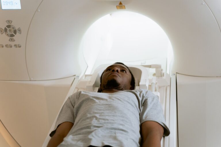
An MRI is typically ordered at the first sign of cognitive problems serious enough to warrant medical evaluation. Guidelines recommend brain imaging for anyone presenting with cognitive complaints or suspected dementia, though not necessarily for every person with normal aging-related forgetfulness. The trade-off is that MRI can be expensive, requires the patient to remain still in a noisy machine for 20 to 45 minutes (which can be distressing for someone with dementia), and takes time to schedule. For practical purposes, if a person’s primary care doctor suspects Alzheimer’s disease, they will order an MRI early in the workup.
In a memory clinic setting, MRI is almost always part of the initial assessment. The urgency depends on the severity of cognitive changes and whether any red flags suggest a treatable condition. Someone with rapid cognitive decline over weeks might need MRI more urgently than someone with slow, gradual decline over years. Once the MRI is done and other conditions are ruled out, it serves as a baseline reference point; a follow-up MRI years later can show whether atrophy has progressed as expected in Alzheimer’s disease, though MRI is not typically repeated frequently for monitoring purposes.
What Are the Key Limitations of MRI in Alzheimer’s Diagnosis?
The most critical limitation is that standard MRI cannot detect the microscopic pathology—the amyloid plaques and tau tangles—that define Alzheimer’s disease at the cellular level. A person could have completely normal-looking brain structure on MRI yet have significant amyloid and tau accumulation showing up on positron emission tomography (PET) scans, a more specialized imaging technique. This means an MRI can miss early Alzheimer’s disease when cognitive changes are minimal but pathological changes are already present. Another limitation is that MRI findings in older adults are often ambiguous.
Many cognitively normal 80-year-olds have hippocampal atrophy indistinguishable from that seen in people with early Alzheimer’s disease. Radiologists cannot reliably tell an MRI that belongs to someone with Alzheimer’s disease from one belonging to a healthy older person just by looking at the images. Additionally, MRI quality and interpretation depend heavily on the skill and experience of the radiologist. A radiologist who specializes in dementia imaging will measure hippocampal volume more accurately than a general radiologist, and different measurement methods can yield different results. Poor image quality due to patient movement or scanner limitations can further compromise accuracy.
Can MRI Distinguish Alzheimer’s Disease From Other Types of Dementia?
MRI patterns can hint at different dementia types but cannot definitively separate them. Alzheimer’s disease typically shows prominent hippocampal and medial temporal lobe atrophy. In contrast, frontotemporal dementia preferentially affects the frontal and anterior temporal lobes, sparing the hippocampus initially. Someone with frontotemporal dementia might have personality changes, language problems, or compulsive behaviors alongside MRI changes that look quite different from Alzheimer’s.
A trained neurologist and radiologist working together can use the atrophy pattern to narrow the differential diagnosis. However, many patients have mixed pathology—Alzheimer’s changes plus vascular disease, for example—that produces overlapping MRI findings. This ambiguity underscores why MRI, while useful, must be paired with clinical assessment, detailed cognitive testing, and increasingly, biomarker testing. The pattern of atrophy can provide helpful context, but it is not a substitute for the full evaluation.
What New MRI Techniques Are Emerging for Better Alzheimer’s Detection?
Advanced MRI techniques like diffusion tensor imaging (DTI) and functional MRI (fMRI) can reveal subtle white matter changes and alterations in brain connectivity that standard structural MRI might miss. These techniques show how different brain regions communicate with each other; in Alzheimer’s disease, those networks become disrupted even before obvious atrophy appears. Research studies using these advanced methods have shown they can sometimes detect early Alzheimer’s changes when standard MRI appears relatively normal.
High-field MRI (using stronger magnetic fields, typically 7 Tesla instead of the standard 1.5 or 3 Tesla) can produce even more detailed images of small structures like the hippocampus, potentially improving early detection. However, these advanced techniques are not yet standard in clinical practice and remain primarily research tools. They are expensive, available only at specialized centers, and their results still must be interpreted in clinical context. As these techniques move from research into clinical use, they may improve the timing and accuracy of Alzheimer’s diagnosis, but they will not replace the need for cognitive testing and other clinical assessment.
Frequently Asked Questions
If my MRI is normal, does that mean I don’t have Alzheimer’s?
A normal MRI does not rule out Alzheimer’s disease, especially in early stages. Some people with Alzheimer’s pathology (amyloid and tau) show minimal brain atrophy on standard MRI. Cognitive testing and biomarker tests provide additional diagnostic information.
How much does an MRI for Alzheimer’s evaluation cost?
Brain MRI typically costs between $1,000 and $2,500 without insurance, depending on the facility and location. Many insurance plans cover it when ordered by a physician for evaluation of cognitive concerns, though the patient may owe a copay or deductible.
Can MRI tell the difference between Alzheimer’s and normal aging?
MRI can show atrophy patterns, but individual variation is high. Many cognitively healthy older adults have brain atrophy similar to people with Alzheimer’s. A radiologist cannot reliably distinguish normal aging from Alzheimer’s on MRI alone without clinical context.
Will I need another MRI if I’m diagnosed with Alzheimer’s?
Follow-up MRI is not routinely ordered unless symptoms change in unexpected ways or to track disease progression in research studies. Most Alzheimer’s care relies on cognitive assessments rather than repeated imaging.
Is MRI safer than other brain imaging tests for dementia?
MRI uses no radiation, making it safer than PET scans or CT scans. However, MRI is not suitable for people with certain metal implants like pacemakers. The procedure involves being still in a loud, enclosed space for 20 to 45 minutes, which some people find claustrophobic or stressful.
Can MRI detect early-stage Alzheimer’s disease?
MRI is most useful for detecting moderate Alzheimer’s disease with obvious atrophy. In very early stages, standard MRI may appear normal even though amyloid and tau pathology is accumulating. PET imaging or blood biomarkers may detect earlier changes.