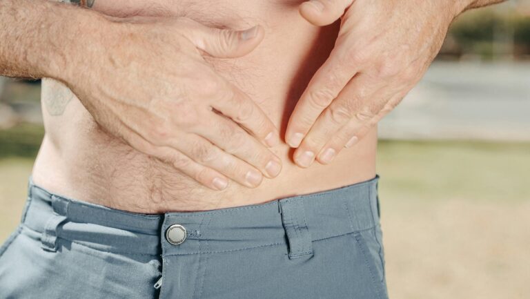
Lumbar spine instability occurs when the vertebrae, discs, and supporting structures of the lower back lose their ability to maintain proper alignment and control during movement. Doctors frequently diagnose eight primary causes: degenerative disc disease, facet joint arthritis, spondylolisthesis, muscle weakness and deconditioning, ligamentous laxity, pars interarticularis defects, poor posture and biomechanical dysfunction, and prior trauma or injury. For example, a 55-year-old patient experiencing chronic lower back pain with radiating symptoms into the buttocks might be diagnosed with facet joint arthritis combined with disc degeneration—a combination that destabilizes the spine and limits normal activities. This article examines each of these eight causes, explains how they develop, and describes what makes them clinically significant for diagnosis and treatment planning.
Table of Contents
- What Is Degenerative Disc Disease and How Does It Cause Instability?
- How Does Facet Joint Arthritis Contribute to Spinal Instability?
- What Role Does Spondylolisthesis Play in Lumbar Instability?
- How Does Muscle Weakness and Deconditioning Destabilize the Spine?
- What Is Ligamentous Laxity and Why Does It Matter?
- What Are Pars Interarticularis Defects and Spondylolysis?
- How Do Postural Problems and Biomechanical Dysfunction Cause Instability?
- Conclusion
- Frequently Asked Questions
What Is Degenerative Disc Disease and How Does It Cause Instability?
Degenerative disc disease (DDD) is the breakdown of the intervertebral discs that cushion the vertebrae. As discs lose water content and structural integrity over time, they become thinner and lose their ability to absorb shock and distribute forces evenly across the spine. When a disc degenerates significantly, the vertebrae above and below it can shift excessively during movement, creating the unstable segment that doctors detect on imaging.
This condition typically develops gradually over decades due to normal aging, though repetitive strain, heavy lifting, or smoking can accelerate the process. However, it’s important to note that disc degeneration on an MRI doesn’t automatically cause instability—many people have degenerative discs without any symptoms. The instability becomes clinically significant only when the degenerative changes are severe enough to allow abnormal movement between vertebrae.

How Does Facet Joint Arthritis Contribute to Spinal Instability?
The facet joints are small joints located on both sides of the spine where vertebrae connect. These joints allow controlled flexion, extension, and rotation. When facet joint arthritis develops—either from aging, repetitive stress, or previous injury—the cartilage surfaces deteriorate and bone spurs may form.
As the joints become damaged, they lose their ability to guide and control motion between vertebrae, allowing increased translation (forward or backward shifting) during movement. A patient with severe facet arthritis might experience increased back pain when standing upright for extended periods because the damaged facet joints cannot properly stabilize the spine against gravity. However, not all facet arthritis causes instability; many patients with mild to moderate facet joint changes maintain adequate stability because their muscles and ligaments compensate effectively. Instability typically develops when facet joint damage is combined with other factors like disc degeneration or muscle weakness.
What Role Does Spondylolisthesis Play in Lumbar Instability?
Spondylolisthesis occurs when one vertebra slips forward or backward relative to the vertebra below it. This condition can develop through several mechanisms: degenerative changes, stress fractures in the pars interarticularis, or structural abnormalities present since birth. Grade 1 spondylolisthesis involves a slip of up to 25 percent of the vertebral body width, while higher grades indicate more severe displacement.
A typical clinical example is a 45-year-old patient with degenerative spondylolisthesis at L4-L5, where years of wear and tear on the facet joints and discs finally cause the L4 vertebra to slip forward, creating a pinched nerve and leg pain when walking (neurogenic claudication). The degree of vertebral slippage directly correlates with spinal instability—the greater the slip, the more excessive movement occurs during daily activities. Treatment decisions depend heavily on the grade of slippage and whether symptoms can be managed conservatively.

How Does Muscle Weakness and Deconditioning Destabilize the Spine?
The deep stabilizing muscles of the core—particularly the multifidus and transverse abdominis—function as a dynamic support system for the spine. When these muscles weaken through inactivity, injury recovery without proper rehabilitation, or chronic pain that causes avoidance of exercise, the spine loses active stabilization. A sedentary office worker with a history of back injury might develop progressive muscle atrophy and weakness, leading to poor control and excessive motion during bending or lifting activities.
This creates a paradoxical situation: weak muscles cannot protect the spine from instability, so movement becomes painful, which further discourages activity and worsens the muscle weakness. The important distinction is that muscle weakness alone may not show up on imaging tests like MRI or X-ray, yet it can be the primary driver of symptomatic instability. Physical therapy focused on core muscle strengthening is often the first-line treatment because restoring muscular support can significantly reduce instability even when structural damage is present.
What Is Ligamentous Laxity and Why Does It Matter?
The spinal ligaments—particularly the anterior longitudinal ligament, posterior longitudinal ligament, and interspinous ligaments—provide passive stabilization by limiting excessive motion between vertebrae. Ligamentous laxity develops when these ligaments are stretched or weakened, either from chronic excessive motion, inflammatory conditions like rheumatoid arthritis, or acute injuries that partially tear the ligament fibers.
An athlete with a history of lumbar strain injuries might develop progressive laxity in the interspinous ligaments from repeated overstretching, resulting in increased segmental motion that causes persistent pain. Unlike muscular stabilization, ligamentous support cannot be quickly restored through exercise—healing tissue cannot fully regain pre-injury elasticity and strength. For patients with significant ligamentous laxity, physical therapy must emphasize muscular compensation strategies to stabilize the spine, since the passive restraint cannot be reliably restored.

What Are Pars Interarticularis Defects and Spondylolysis?
The pars interarticularis is a thin bridge of bone connecting the upper and lower facet joints on each side of the vertebra. A stress fracture in this bone structure creates a defect called spondylolysis, which most commonly occurs at the L5 vertebra. This condition develops from repetitive hyperextension and rotation, which is why it’s frequently seen in athletes participating in gymnastics, weightlifting, or football.
When a unilateral pars defect (fracture on one side) occurs without slippage, stability may be maintained adequately through muscle support and the remaining structures. However, when bilateral pars defects occur or the fracture progresses to cause spondylolisthesis, significant instability develops because the mechanical linkage between the vertebral bodies is compromised. Doctors distinguish between acute spondylolysis (a recent fracture) and chronic spondylolysis (a healed defect with scar tissue), as chronic defects are more likely to cause long-term instability.
How Do Postural Problems and Biomechanical Dysfunction Cause Instability?
Poor posture and abnormal movement patterns place repetitive stress on specific spine structures, accelerating the development of degenerative changes and creating functional instability. A person who habitually rounds their lower back while sitting, stands with excessive anterior pelvic tilt, or moves using upper back muscles instead of core muscles may experience progressive wear and stress on discs and facet joints.
For example, a patient who consistently lifts heavy objects using their back instead of their legs places enormous shear forces on the lumbar spine with each repetition, potentially causing disc herniation and accelerating disc degeneration. The challenging aspect of biomechanical dysfunction is that it’s often deeply ingrained through years of habit—correcting poor movement patterns requires conscious effort and practice over weeks and months. However, improving biomechanics through physical therapy can often prevent further deterioration and reduce symptoms even when underlying structural damage is already present.
Conclusion
Lumbar spine instability results from multiple interacting factors rather than a single cause. The eight causes that doctors most frequently diagnose—degenerative disc disease, facet joint arthritis, spondylolisthesis, muscle weakness, ligamentous laxity, pars interarticularis defects, postural dysfunction, and prior trauma—often occur together in varying combinations. Understanding which specific causes contribute to a patient’s instability guides treatment decisions, from conservative approaches emphasizing muscle strengthening and movement correction to surgical stabilization for severe cases.
If you experience persistent lower back pain, instability sensation (feeling like your back is “giving out”), or pain that worsens with prolonged standing or activity, consult a spine specialist for proper evaluation. Modern diagnostic imaging combined with physical examination can identify the specific causes of instability, allowing for targeted treatment that addresses the underlying dysfunction rather than simply managing symptoms. Early intervention to restore muscle strength and address biomechanical issues often prevents progression to more severe instability.
Frequently Asked Questions
Can lumbar spine instability be cured without surgery?
Many patients achieve significant improvement through conservative treatment including physical therapy, core strengthening, postural correction, and activity modification. Surgery is typically reserved for severe cases with neurological compromise or when conservative treatment fails after 3-6 months. Success depends on addressing the underlying causes rather than expecting a single treatment to resolve instability.
How do doctors determine which specific cause is responsible for my instability?
Diagnosis combines imaging studies (MRI, X-ray, sometimes CT scanning), physical examination testing for abnormal movement patterns, muscle strength assessment, and patient history. A skilled clinician integrates all this information to identify which of the eight common causes applies to each patient.
Is lumbar instability permanent?
Structural damage like spondylolisthesis or severe disc degeneration cannot be reversed, but the symptoms and functional limitations caused by instability can often be significantly improved through appropriate management. The goal is functional stability through muscle support rather than anatomical restoration.
Why does my pain sometimes improve without any specific treatment?
The inflammatory phase of injury naturally improves over weeks to months as swelling decreases. Additionally, your body may unconsciously stabilize the unstable segment through muscle guarding, though this often creates secondary problems like muscle tension and reduced mobility.
Can I exercise with lumbar spine instability?
Appropriate exercise is essential for managing instability, but the type of exercise matters significantly. Core stabilization exercises and proper movement patterns help, while heavy lifting, hyperextension movements, or unstable exercises like certain yoga positions may worsen symptoms. Work with a physical therapist to develop an appropriate program.