Pelvic instability in adults stems from seven well-documented risk factors: pregnancy and childbirth, trauma and injury, advancing age and menopause, obesity, prior pelvic surgery, connective tissue disorders, and athletic overuse with muscle imbalances. Any one of these can compromise the ligaments, muscles, and bony structures that hold the pelvic ring together, but many adults live with two or three overlapping risk factors without realizing it. Consider a 65-year-old woman who had two difficult deliveries decades ago, went through menopause at 52, and now carries extra weight around her midsection. She may assume her chronic lower back pain and bladder leakage are just aging, when in fact her pelvis has been gradually losing structural integrity for years across multiple fronts. The numbers behind pelvic instability deserve attention.
According to research published in Nature Scientific Reports, 32% of women have at least one pelvic floor disorder, and over 50% of women past age 80 are affected. Projections suggest the number of U.S. women dealing with pelvic floor disorders will climb from 28.1 million in 2010 to 43.8 million by 2050, a 56% increase driven largely by an aging population. For families navigating dementia care, pelvic instability adds another layer of complexity, since falls, immobility, and incontinence are already major concerns in that population. This article breaks down each of the seven risk factors, explains how they interact, and offers practical guidance for reducing risk before instability takes hold.
Table of Contents
- What Are the Primary Risk Factors That Lead to Pelvic Instability in Adults?
- How Pregnancy and Childbirth Reshape Pelvic Stability for Years
- The Compounding Effect of Trauma, Falls, and Aging on Pelvic Integrity
- Managing Weight and Surgical History to Protect Pelvic Stability
- Why Connective Tissue Disorders Make Pelvic Instability Harder to Predict and Treat
- Athletic Overuse and the Muscle Imbalances That Destabilize the Pelvis
- The Growing Scale of Pelvic Instability and What Lies Ahead
- Conclusion
- Frequently Asked Questions
What Are the Primary Risk Factors That Lead to Pelvic Instability in Adults?
The pelvis is not a single bone but a ring of bones held together by some of the strongest ligaments in the body. When those ligaments stretch, tear, or weaken, the ring loses its structural tension and the joints begin to move in ways they should not. The seven risk factors discussed here all attack this system through different mechanisms. Pregnancy floods the body with relaxin, a hormone that deliberately loosens pelvic ligaments. Trauma can tear them outright. Age and hormonal changes erode their elasticity over decades. Obesity loads them beyond their capacity.
Surgery disrupts them mechanically. Connective tissue disorders mean they were never as strong as they should have been in the first place. And athletic overuse grinds them down through repetition. What makes pelvic instability particularly tricky is that these risk factors are cumulative. A young woman with Ehlers-Danlos syndrome who then becomes pregnant faces a far greater risk than either factor alone would suggest. Similarly, a man who sustained a pelvic fracture in a car accident at 30 may do fine for two decades, only to develop chronic instability in his fifties as age-related muscle loss removes the compensatory support his body had been providing. Compared to a joint like the knee, where instability tends to produce obvious symptoms quickly, pelvic instability often develops gradually, and people learn to accommodate it unconsciously by changing how they walk, sit, or stand, which creates secondary problems in the hips and lower back.

How Pregnancy and Childbirth Reshape Pelvic Stability for Years
The placenta secretes relaxin specifically to increase flexibility of the pelvic ligaments and soften the cervix, preparing the body for delivery. This is necessary and normal, but it comes at a cost. The pelvic joints, particularly the pubic symphysis at the front and the sacroiliac joints at the back, become measurably less stable during pregnancy. For most women, this resolves within months of delivery. For some, it does not. Specific obstetric risk factors that increase the likelihood of lasting pelvic instability include first pregnancy, cephalopelvic disproportion where the baby is large relative to the mother’s pelvic capacity, breech presentation, prolonged or traumatic labor, twin pregnancy, and instrumented delivery using forceps or ventouse.
However, it is important not to overstate this risk. The majority of women who give birth, even those with difficult deliveries, recover full pelvic stability. The women who do not tend to have additional overlapping risk factors: pre-existing hypermobility, insufficient postpartum rehabilitation, or a return to heavy physical activity before the ligaments have fully recovered their tension. A critical limitation of the research in this area is that many studies focus on pelvic floor dysfunction (incontinence and prolapse) rather than pelvic ring instability specifically. These are related but distinct problems. A woman can have a stable pelvic ring but weak pelvic floor muscles, or vice versa. Treatment for one does not automatically address the other, which is why a proper clinical assessment matters more than assumptions based on birth history alone.
The Compounding Effect of Trauma, Falls, and Aging on Pelvic Integrity
Prior traumatic disruptions to the pelvic ring, from falls, motor vehicle accidents, or sports injuries, are a documented cause of chronic anterior pelvic instability, particularly when the original injury was managed non-operatively. This matters enormously in the context of older adults and dementia care. A person who fractured their pelvis in a fall five years ago may have healed the bone but never fully restored the ligament integrity. Now, as they age and potentially develop cognitive decline, their balance deteriorates, their muscle mass drops, and the pelvis that was barely holding together becomes a source of chronic pain and functional limitation. Age itself is the single most significant risk factor for pelvic floor disorders.
The Nature Scientific Reports study found that over 50% of women over 80 have at least one pelvic floor disorder. Menopausal hormonal changes accelerate the weakening of pelvic support structures by reducing the collagen content in ligaments and connective tissue. For men, age-related sarcopenia, the loss of muscle mass, strips away the muscular scaffolding that compensates for ligament laxity. A specific example illustrates the danger: an 82-year-old man with moderate Alzheimer’s who sustained a pelvic ring injury from a bathroom fall may not be able to reliably report his pain or comply with rehabilitation exercises. His caregivers might attribute his reluctance to walk to his dementia rather than to an unstable pelvis, leading to further deconditioning and a downward spiral of immobility.

Managing Weight and Surgical History to Protect Pelvic Stability
Higher BMI is strongly and significantly associated with all three categories of pelvic floor disorder: urinary incontinence, pelvic organ prolapse, and bowel dysfunction. The mechanism is straightforward. Excess abdominal weight increases the chronic downward pressure on the pelvic floor and the outward pressure on the pelvic ring. Repeated heavy lifting, whether occupational or recreational, compounds this effect. The tradeoff that many people face is that exercise is one of the best ways to strengthen the muscles that support the pelvis, but certain types of exercise, particularly heavy barbell movements, temporarily increase intra-abdominal pressure and can worsen instability in someone already at risk. The practical compromise is to prioritize exercises that build pelvic stability without excessive loading: bodyweight movements, resistance bands, swimming, and targeted pelvic floor training, while working toward a healthier weight over time.
Prior pelvic surgery is the other risk factor in this pair, and it interacts with weight in important ways. Hysterectomy and other urological or gynecological interventions are documented risk factors for pelvic instability because they disrupt the connective tissue architecture that holds the pelvic organs in position. A woman who has had a hysterectomy and then gains significant weight faces a double challenge: the surgical disruption reduced her structural reserves, and the added weight is now exceeding what remains. Compared to someone with no surgical history, she has less margin for error. Adequate rehabilitation after pelvic surgery is critical, yet it is frequently underprescribed. Many patients receive instructions to avoid heavy lifting for six weeks and then are discharged without any structured pelvic rehabilitation program.
Why Connective Tissue Disorders Make Pelvic Instability Harder to Predict and Treat
Genetic conditions like Ehlers-Danlos syndrome and hypermobility spectrum disorders significantly increase susceptibility to pelvic instability because the collagen that forms the structural basis of ligaments is inherently weaker or more elastic than normal. Hypermobility syndrome is listed as a known etiology for chronic anterior pelvic ring instability. The challenge with these conditions is that standard treatment protocols, designed for people with normal connective tissue, may not work. Ligament-tightening exercises can help a person with normal collagen regain pelvic stability, but for someone with a connective tissue disorder, those same exercises may have limited effect because the ligaments will never develop normal tension. A warning for clinicians and caregivers: connective tissue disorders are frequently undiagnosed or underdiagnosed, particularly in older adults.
A person who was “always flexible” and had “loose joints” their whole life may carry an unrecognized hypermobility spectrum disorder into their seventies. When that person develops pelvic instability, it may be attributed entirely to age, and the underlying connective tissue problem goes unaddressed. This matters for treatment planning because these individuals often need external support, bracing, or surgical intervention sooner than the general population. They also have higher rates of surgical complications and slower healing, which limits the available options. Screening for hypermobility should be part of any pelvic instability workup, but in practice it is frequently overlooked, especially in elderly patients whose joint range of motion has decreased with age, masking the underlying hypermobility.

Athletic Overuse and the Muscle Imbalances That Destabilize the Pelvis
Intense physical activity and overuse injuries can cause degenerative conditions including osteitis pubis, a painful inflammation of the pubic symphysis that serves as a pathway to pelvic instability. This is most commonly seen in runners, soccer players, and hockey players who subject the pelvis to repetitive asymmetric loading. But the risk extends beyond elite athletes. A weekend warrior who runs 30 miles a week while neglecting core strengthening can develop the same muscle imbalances.
Specifically, weakening of the transverse and oblique abdominal muscles reduces pelvic support and increases instability risk. These deep core muscles act as a corset around the pelvis, and when they are overpowered by stronger hip flexors and spinal extensors, the pelvis tilts and rotates in ways that stress the ligaments beyond their capacity. For older adults, the athletic overuse risk factor may seem irrelevant, but it manifests differently. A retiree who takes up gardening, spending hours bent over in asymmetric positions, or a caregiver who repeatedly lifts a family member with dementia using poor body mechanics, can develop the same muscle imbalances and pelvic stress that affect younger athletes. The underlying principle is the same: repetitive, asymmetric loading without adequate muscular support destabilizes the pelvis over time.
The Growing Scale of Pelvic Instability and What Lies Ahead
The epidemiological trajectory is clear. Urinary incontinence alone is projected to rise from 18.3 million affected U.S. women to 28.4 million by 2050, a 55% increase. These projections reflect demographic realities, an aging population with increasing rates of obesity, that are not going to reverse quickly. For dementia care specifically, pelvic instability represents an under-recognized contributor to falls, incontinence, and reduced mobility, three problems that already dominate the caregiving landscape.
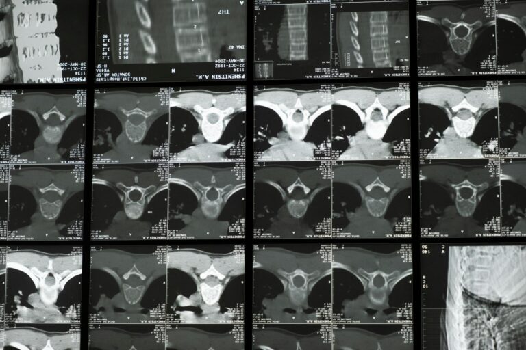
As awareness grows, there is reason to hope that screening for pelvic instability will become more routine in geriatric assessments, and that rehabilitation programs will be designed to address the specific combination of risk factors each individual carries rather than treating pelvic instability as a single condition with a one-size-fits-all response. The most promising developments are in early identification and multimodal rehabilitation. Imaging techniques have improved to the point where subtle pelvic ring instability can be detected before it becomes symptomatic. Pelvic floor physical therapy, once considered niche, is now widely available and increasingly covered by insurance. And there is growing recognition among orthopedic and gynecological specialists that pelvic instability requires a team approach rather than a single-specialty solution. For caregivers supporting someone with cognitive decline, the practical takeaway is to advocate for pelvic assessment whenever unexplained pain, gait changes, or worsening incontinence appear, rather than assuming these are inevitable consequences of aging or dementia.
Conclusion
Pelvic instability in adults rarely has a single cause. The seven risk factors, pregnancy and childbirth, trauma, aging and menopause, obesity, prior surgery, connective tissue disorders, and athletic overuse, frequently overlap and compound each other in ways that make the condition both more likely and harder to treat. Understanding which risk factors apply to a specific individual is the first step toward meaningful prevention and management. For the 32% of women already living with at least one pelvic floor disorder, and for the millions of older adults whose pelvic stability is quietly eroding, awareness of these risk factors is not academic.
It is the difference between proactive intervention and reactive crisis management. If you or someone you care for has multiple risk factors on this list, bring them to the attention of a healthcare provider who can assess pelvic stability directly. Pelvic floor physical therapy, weight management, targeted strengthening of the deep core muscles, and appropriate bracing or surgical options can all make a real difference, but only if the problem is recognized. In the context of dementia care, where communication barriers make self-reporting unreliable, caregivers play a critical role in watching for the signs: changes in gait, new or worsening incontinence, reluctance to move, and unexplained pain behaviors. Early assessment and intervention can preserve mobility, reduce fall risk, and maintain quality of life in ways that benefit both the person affected and those who care for them.
Frequently Asked Questions
Can pelvic instability be fully reversed, or is it a permanent condition?
It depends on the cause and severity. Pregnancy-related pelvic instability often resolves with time and rehabilitation. Instability caused by connective tissue disorders like Ehlers-Danlos syndrome cannot be fully reversed but can be managed with targeted physical therapy, bracing, and in some cases surgery. Trauma-related instability falls somewhere in between, with outcomes depending on the extent of the original injury and the quality of rehabilitation.
How is pelvic instability different from pelvic floor dysfunction?
Pelvic instability refers to abnormal movement in the bony pelvic ring and its ligaments, while pelvic floor dysfunction involves weakness or dysfunction of the muscles that span the bottom of the pelvis. They frequently coexist and share many of the same risk factors, but they require different assessments and sometimes different treatments. A person can have one without the other.
Does pelvic instability increase fall risk in people with dementia?
Yes. Pelvic instability compromises balance and gait stability, which adds to the fall risk already elevated by cognitive decline, medication side effects, and environmental factors common in dementia. Because people with dementia may not be able to articulate that their pelvis hurts or feels unstable, caregivers should watch for behavioral cues like reluctance to stand, shuffling gait, or guarding one side of the body.
At what age should screening for pelvic instability begin?
There is no universal guideline, but risk-based screening makes sense. Women after childbirth, anyone after a pelvic injury, and all adults over 60 with symptoms like incontinence, lower back pain, or gait changes should be evaluated. For individuals with known connective tissue disorders, monitoring should begin earlier and continue throughout life.
Can men develop pelvic instability, or is it primarily a female condition?
Men can and do develop pelvic instability, though it is less common than in women due to the absence of pregnancy-related risk and the generally narrower, more inherently stable male pelvis. Male pelvic instability most often results from trauma, athletic overuse (particularly osteitis pubis), prior surgery, or age-related muscle loss. It is underdiagnosed in men partly because clinicians may not consider it.