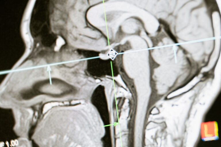
FDG-PET scans reveal Alzheimer’s disease by showing where the brain is struggling to use glucose for energy. The scan creates a color-coded map of brain metabolism: areas that appear dimmer—showing reduced glucose uptake—are regions where nerve cells aren’t firing properly, a pattern called hypometabolism. In a healthy brain, energy use is relatively uniform across regions. In Alzheimer’s, the scan typically shows a distinctive dimming in the temporoparietal cortex (the area where the temporal and parietal lobes meet), along with the precuneus and posterior cingulate.
This metabolic breakdown happens because dying neurons and deteriorating synapses simply can’t maintain normal energy consumption, even if the brain tissue itself still appears intact on a standard MRI scan. What makes FDG-PET unique is its ability to detect this metabolic failure before cognitive decline becomes obvious. A person might pass standard memory tests yet show clear hypometabolism on their FDG-PET scan—a signal that neurodegeneration is already underway. This early warning capability has made the scan increasingly valuable in diagnosing mild cognitive impairment and predicting who will progress to dementia, which is why major medical centers now use it as a standard diagnostic tool alongside MRI and cognitive testing.
Table of Contents
- How FDG-PET Detects Brain Metabolism Changes in Alzheimer’s
- The Classic Hypometabolism Pattern in Alzheimer’s Disease
- Diagnostic Accuracy Compared to Other Brain Imaging
- Early Detection Before Cognitive Decline Becomes Noticeable
- When FDG-PET Patterns Differ and What That Means
- Recent Clinical Advances and Emerging Applications
- Insurance Coverage and Access Considerations
How FDG-PET Detects Brain Metabolism Changes in Alzheimer’s
The scan works by injecting a tracer—typically a glucose molecule tagged with a radioactive fluorine isotope, called fluorodeoxyglucose or FDG—into the patient’s bloodstream. Brain cells that are metabolically active consume more glucose, so they accumulate more of the tracer. A specialized camera then maps where that radiation is concentrated, creating an image that directly reflects which brain regions are working hardest. The tracer doesn’t show physical structure the way an mri does; instead, it shows *function*—how much energy each area is burning moment to moment. Neurologists refer to areas of reduced glucose use as hypometabolism. In Alzheimer’s disease, hypometabolism reflects the loss of synaptic connections and the death of neurons in those regions.
A cell that’s degenerating or disconnected from its network simply doesn’t need as much fuel. For comparison, a stroke causes a sudden, sharp drop in blood flow and glucose use in a well-defined region, producing a stark boundary on the scan. Alzheimer’s hypometabolism is usually more gradual and spreads across multiple interconnected brain areas, reflecting the diffuse nature of the disease’s neuronal loss. One important caveat: the presence of hypometabolism doesn’t automatically mean a patient has Alzheimer’s. The scan must be interpreted in context of the person’s symptoms, cognitive testing results, and other imaging. Depression, for instance, can produce metabolic changes in certain brain regions, and other dementias like frontotemporal dementia show different patterns. That’s why FDG-PET is most useful as one piece of the diagnostic puzzle, not as a standalone test.
The Classic Hypometabolism Pattern in Alzheimer’s Disease
The hallmark FDG-PET pattern in Alzheimer’s is bilateral hypometabolism affecting the temporoparietal cortex—both sides of the brain’s outer layer where the temporal and parietal lobes meet. This area sits behind and above the ears and extends toward the back of the head. Additional hypometabolism typically shows up in the precuneus (deep inside the brain, part of the default mode network) and the posterior cingulate (a band of tissue just above the back of the corpus callosum). Together, these regions form what radiologists call the “Alzheimer’s pattern,” and it appears in studies with sensitivity of 74.2% to 93.6% depending on which specific region you’re measuring and how strict the criteria are. The variability matters clinically.
Some patients show pronounced temporoparietal hypometabolism but only mild changes in the posterior cingulate; others show the opposite. A 2025-2026 longitudinal study tracking 1,136 cognitively normal people over ten years found that the specific regions affected on early FDG-PET scans could actually predict which disease pathway a person would follow—some progressing rapidly, others slowly, still others not progressing at all over the study period. This suggests that the exact topography of hypometabolism early on contains information about future disease trajectory, not just whether disease is present. A significant limitation is that hypometabolism can precede actual cognitive symptoms by years or even decades. Some cognitively normal older adults show Alzheimer’s-pattern hypometabolism on FDG-PET but never develop dementia during their remaining lifespan. This creates a diagnostic dilemma: should asymptomatic people with this finding be told they have “preclinical Alzheimer’s,” and if so, should they undergo preventive treatment? Medical guidelines continue to evolve on this question, and the answer depends partly on whether the person carries genetic risk factors like APOE4.
Diagnostic Accuracy Compared to Other Brain Imaging
When evaluated against cognitive decline and pathological confirmation of Alzheimer’s disease, FDG-PET shows solid diagnostic accuracy. Studies using standard visual assessment (looking at the scan images) report accuracy around 84.8%, while computer-assisted measurement using stereotactic surface projection reaches 89.2%. But here’s where recent technology is changing the picture: artificial intelligence algorithms trained to recognize Alzheimer’s patterns on FDG-PET images achieve what’s called an SROC-AUC (Summary Receiver Operating Characteristic Area Under the Curve) of 0.96 with a 95% confidence interval of 0.94 to 0.98. That makes AI-assisted FDG-PET slightly better than structural MRI, which scores 0.94 on the same scale. This AI advantage reflects a fundamental difference in what each modality sees. An MRI excels at showing brain shrinkage—widened ventricles, thinned cortex, hippocampal atrophy—which are structural markers of neurodegeneration.
FDG-PET shows the *functional* consequence of that neurodegeneration: the loss of metabolic activity in the affected cells. In some early-stage cases, a patient might show hypometabolism on FDG-PET before they show measurable brain atrophy on MRI, making the metabolic scan sometimes more sensitive in catching the disease early. The catch is that FDG-PET is far more expensive and time-consuming than an MRI or a cognitive test battery. A single FDG-PET scan can cost $3,000 to $5,000 depending on the facility, and it requires the patient to remain still in the scanner for 20 to 30 minutes after the tracer injection. Most insurance plans, including Medicare, now cover it for dementia workup—but only when ordered by a neurologist or geriatrician as part of a formal diagnostic evaluation. Screening asymptomatic older adults or using it to monitor disease progression over time remains uncommon due to cost and radiation exposure.
Early Detection Before Cognitive Decline Becomes Noticeable
One of the most significant clinical roles for FDG-PET is identifying people with mild cognitive impairment (MCI) who are likely to progress to dementia. In MCI, the person notices subtle memory or thinking changes and performs below expected on cognitive testing, but these changes don’t yet interfere significantly with daily function. FDG-PET hypometabolism in the Alzheimer’s pattern predicts progression: in one two-year follow-up study, sensitivity for progression was 70% and specificity was 90%, meaning the scan correctly identified most people who would decline while correctly identifying most who would remain stable. The timeline of metabolic change is important. Hypometabolism can appear in cognitively normal people—those with no noticed thinking changes and normal cognitive testing—who carry amyloid or tau pathology in their brains.
A 10-year longitudinal study of cognitively normal participants found that baseline FDG-PET glucose hypometabolism predicted which individuals would later develop cognitive impairment, even accounting for amyloid status. This suggests that metabolic failure is part of the disease cascade and adds predictive information beyond what amyloid or tau biomarkers alone provide. However, predicting future decline in an asymptomatic person raises ethical questions that individual clinicians must navigate with their patients. Some centers now offer research studies where cognitively normal older adults with amyloid positivity and hypometabolism on FDG-PET are offered experimental disease-modifying treatments, similar to how clinical trials evaluate early intervention. But for most patients and clinicians, FDG-PET remains most useful once mild cognitive impairment is already present, helping confirm that the MCI is due to Alzheimer’s pathology rather than depression, medication effects, or other causes.
When FDG-PET Patterns Differ and What That Means
Not all dementia looks the same on FDG-PET, and this is where the scan becomes genuinely diagnostic rather than merely supportive. In frontotemporal dementia (FTD), hypometabolism concentrates heavily in the frontal and anterior temporal lobes, sparing the posterior regions affected in Alzheimer’s. The scan can differentiate Alzheimer’s from FTD with 99% sensitivity and 65% specificity in some studies—meaning it’s extremely good at recognizing when the pattern is *not* typical Alzheimer’s, though it has more trouble ruling in FTD uniquely. Dementia with Lewy bodies (DLB) produces a different metabolic signature: relative preservation of the posterior temporal lobe but hypometabolism in the occipital cortex (the brain’s vision center) and parietal regions. A patient with DLB might report visual hallucinations or movement problems as their primary symptom, and the FDG-PET pattern can support that diagnosis.
Vascular dementia tends to show more patchy, regional hypometabolism reflecting the territories of blocked or damaged blood vessels, rather than the symmetric bilateral pattern of Alzheimer’s. One important limitation: FDG-PET can be falsely reassuring. Some older adults with cognitive decline show minimal hypometabolism even though they have documented amyloid and tau pathology in their brains. This discordance might reflect differences in how aggressively each individual’s brain compensates for early pathology, or it might suggest that metabolic imaging is simply less sensitive than biomarker testing in preclinical stages. Additionally, patients on certain medications (particularly sedatives or anti-anxiety drugs taken before the scan) can show artificially reduced glucose uptake, requiring either medication adjustment or repeat scanning.
Recent Clinical Advances and Emerging Applications
In May 2026, a new clinical trial launched (NCT07611357) testing [18F]F-AraG PET imaging—a different kind of PET scan designed to visualize T cells and immune activity in Alzheimer’s brains. This represents a shift from measuring neuronal metabolism alone to understanding the inflammatory component of the disease. If validated, this imaging could allow clinicians to track whether new anti-inflammatory or immunotherapy treatments are actually reducing brain inflammation in individual patients, moving beyond the static anatomical picture that current FDG-PET provides.
AI interpretation of FDG-PET has accelerated over the past two years. Software now available at major academic medical centers can automatically identify and quantify hypometabolism in the Alzheimer’s pattern, reducing operator variability and potentially catching subtle changes that a radiologist’s eye might miss on first review. Some of these systems provide a percentage confidence score—essentially telling the ordering physician “this scan is 94% consistent with Alzheimer’s pattern”—which helps standardize communication between radiology and neurology services.
Insurance Coverage and Access Considerations
Medicare coverage decision CAG-00088N approves FDG-PET imaging for patients with cognitive impairment suspected to be due to Alzheimer’s disease when clinically indicated. Most major private insurance plans follow Medicare’s lead, though specific coverage rules vary by plan and region. Coverage typically requires a referring physician (neurologist, geriatrician, or primary care doctor) to document that the patient has cognitive symptoms or testing abnormalities, and that FDG-PET will change management—not merely confirm a diagnosis already suspected on clinical grounds.
Out-of-pocket costs for the uninsured or underinsured typically range from $3,000 to $5,000, though prices vary significantly by geographic region and facility type. Some academic research centers and teaching hospitals offer FDG-PET at reduced rates or free for participants in research studies. When exploring access, it’s worth checking whether your local facility participates in any investigational studies for early Alzheimer’s detection or disease-modifying therapy trials; these often include imaging at no cost to participants. Conversely, some insurance plans explicitly deny FDG-PET for asymptomatic screening or for tracking disease progression in established dementia, reserving coverage for the diagnostic workup phase alone.