Hippocampal atrophy on MRI—the visible shrinkage of a walnut-sized brain structure deep inside the temporal lobe—matters because it’s often the first physical sign that memory loss is rooted in Alzheimer’s disease or another degenerative process, not normal aging or stress. When radiologists measure the hippocampus on a brain scan, they’re looking for loss of volume that suggests the nerve cells in this region have begun to die.
This atrophy doesn’t cause Alzheimer’s disease, but it frequently accompanies the pathological changes—amyloid plaques and tau tangles—that damage neurons and erode memory function over months and years. For patients presenting with memory complaints, a hippocampus that measures smaller than expected for age and brain size can be the imaging finding that tips a diagnosis from “possible cognitive impairment” toward “probable Alzheimer’s disease.” A 65-year-old woman who has been forgetting recent conversations and losing her place in familiar activities might have an MRI that shows her hippocampus is shrunken to half the typical volume—a red flag that neurodegeneration has likely begun. Yet atrophy is not a death sentence; it’s a measurement that helps clinicians understand what is happening and what to expect, and it opens the door to treatments or lifestyle interventions that may slow decline.
Table of Contents
- What Does Hippocampal Atrophy Look Like on an MRI Scan?
- Why Does the Hippocampus Shrink in Dementia?
- The Connection Between Hippocampal Atrophy and Alzheimer’s Disease
- How Do Clinicians Interpret Hippocampal Atrophy in the Clinic?
- What Hippocampal Atrophy Cannot Tell Us—And Why This Matters
- Early Detection and Biomarkers—Why Radiologists Are Measuring More Carefully Now
- A Clinical Scenario: What Atrophy Means in Practice
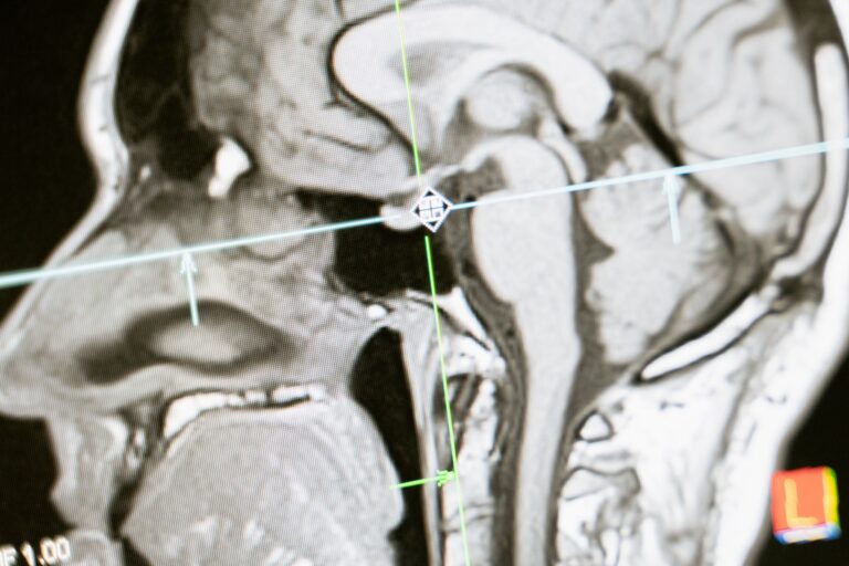
What Does Hippocampal Atrophy Look Like on an MRI Scan?
The hippocampus appears on MRI as a small, curved structure with a distinctive seahorse-like shape—hence its name, from the Greek words for “horse” and “sea.” On a healthy brain MRI, it should have clear borders, smooth contours, and a consistent volume that radiologists can measure using specialized software. When atrophy occurs, that structure shrinks: its borders become less defined, its volume diminishes, and the signal intensity on the scan may change. A radiologist comparing a patient’s current scan to a prior one from several years earlier may see progressive shrinkage, a sign that the atrophy is ongoing rather than static or longstanding.
Measuring hippocampal volume is not eyeballing—it requires tracing the boundaries of the structure on multiple slices of the MRI and calculating the total volume in cubic centimeters. Normal hippocampal volume varies by age, sex, and overall brain size, and radiologists use reference ranges to determine whether a measurement is within expected limits or abnormally small. A 70-year-old man with mild age-related shrinkage might have a hippocampal volume of 2.8 cubic centimeters on each side, while a 70-year-old woman with Alzheimer’s disease might measure 1.9 cubic centimeters—a difference that suggests more than normal aging and points toward neurodegeneration.
Why Does the Hippocampus Shrink in Dementia?
The hippocampus is vulnerable to Alzheimer’s pathology because its neurons depend heavily on a signaling molecule called acetylcholine and because amyloid and tau proteins accumulate there early in the disease process. As plaques and tangles build up, they trigger inflammation, oxidative stress, and ultimately neuronal death—the cells literally die and are cleared away, leaving less tissue behind. The atrophy is the end result of a cascade that may have been unfolding for years before a person notices memory problems.
However, hippocampal shrinkage is not unique to Alzheimer’s disease. It can occur in vascular dementia, when repeated small strokes reduce blood flow to the hippocampus; in Lewy body dementia, where alpha-synuclein protein accumulation damages the structure; and even in conditions unrelated to dementia, such as chronic seizure disorders, severe depression, or prolonged stress. A patient with a 20-year history of poorly controlled temporal lobe epilepsy may have significant hippocampal atrophy from repeated seizures, even without any cognitive impairment. This means that atrophy on its own is a sign of damage, but it does not automatically tell clinicians which disease is responsible.
The Connection Between Hippocampal Atrophy and Alzheimer’s Disease
The hippocampus is one of the first brain regions affected in Alzheimer’s disease because it is the gateway for forming new memories—it tags incoming information and sends it to the cortex for long-term storage. When hippocampal neurons die, people struggle to encode new experiences, which is why memory loss is often the earliest symptom. Autopsies of Alzheimer’s patients consistently show that the hippocampus has accumulated more amyloid and tau than almost any other brain region, and MRI studies show that people with mild cognitive impairment who also have hippocampal atrophy are much more likely to progress to Alzheimer’s dementia within three to five years than those with normal hippocampal volume.
Yet not everyone with a shrunken hippocampus develops Alzheimer’s disease, and not everyone who develops Alzheimer’s shows obvious atrophy early on. Some people accumulate amyloid and tau plaques for years before their hippocampus visibly shrinks on MRI—the pathology exists but the structural damage has not yet reached the point of measurable volume loss. Others may have mild atrophy that plateaus and never causes dementia, especially if they are genetically resilient or lifestyle factors like cognitive stimulation and physical activity provide some protection.
How Do Clinicians Interpret Hippocampal Atrophy in the Clinic?
When a neurologist or geriatrician receives an MRI report noting hippocampal atrophy, they view it as one piece of evidence among many: the patient’s cognitive test scores, their reported symptoms, their family history, and other imaging findings like cortical thickness, white matter disease, or vascular changes. A patient with mild memory complaints, low scores on the Montreal Cognitive Assessment, and moderate bilateral hippocampal atrophy is more likely to receive an Alzheimer’s diagnosis than a cognitively normal patient with incidental atrophy found by chance on an MRI ordered for headaches. The pattern matters—atrophy that is symmetric (equal on both sides) and limited to the hippocampus points more toward Alzheimer’s, while atrophy concentrated in one temporal lobe might suggest a tumor or stroke in the past.
Clinicians also consider rate of change. If an MRI from two years ago showed normal hippocampal volume and a new scan shows clear atrophy, that rapid decline suggests an ongoing neurodegenerative process. In contrast, an MRI showing the same moderate atrophy as a study from a decade earlier suggests the damage may have plateaued, and the cognitive symptoms the patient is experiencing might be due to something else—fatigue, medication side effects, or depression—rather than progressive Alzheimer’s disease.
What Hippocampal Atrophy Cannot Tell Us—And Why This Matters
One critical limitation is that hippocampal atrophy does not predict cognitive impairment reliably at the individual level. A 75-year-old with significantly shrunken hippocampi might have perfect memory and sharp thinking, especially if they have high cognitive reserve from education, lifelong learning, or occupation. Conversely, a person with normal hippocampal volume on MRI but severe cognitive impairment might be experiencing Alzheimer’s pathology that has not yet caused visible structural change, or they might have a different cause of dementia such as frontotemporal dementia, which affects the front of the brain more than the hippocampus. Using atrophy alone to predict who will develop cognitive problems is unreliable and can lead to unnecessary worry or false reassurance.
Another limitation is that MRI scans measure structure, not function. A shrunken hippocampus tells you that neurons have died, but it does not tell you whether the remaining neurons are still firing, whether the circuits connecting the hippocampus to other brain regions are preserved, or whether compensatory changes elsewhere in the brain are supporting memory function. Advanced imaging techniques like PET scans for amyloid and tau, or functional MRI to map connectivity, provide additional information but are not routine clinical tools. For most patients in a primary care or memory clinic setting, the hippocampal measurement is a clue, not a diagnosis.
Early Detection and Biomarkers—Why Radiologists Are Measuring More Carefully Now
Over the past decade, there has been growing emphasis on detecting Alzheimer’s pathology before symptoms appear, using biomarkers like amyloid PET, tau PET, and blood tests that measure phosphorylated tau and amyloid beta. Hippocampal atrophy is considered a “neurodegeneration” biomarker—it shows that brain damage has occurred, but it appears later in the disease cascade than amyloid accumulation.
This means that by the time someone has obvious hippocampal shrinkage on MRI, Alzheimer’s pathology has been building for years, possibly decades. Some research centers now use quantitative hippocampal volume measurements as part of a comprehensive biomarker assessment in patients with mild cognitive impairment, alongside blood tests and other imaging, to stage the degree of neurodegeneration and predict rates of decline.
A Clinical Scenario: What Atrophy Means in Practice
Consider a 72-year-old man with a three-year history of increasing forgetfulness—he repeats stories, forgets appointments despite reminders, and sometimes cannot recall what he ate for lunch. His daughter brought him to a neurologist, who gave him a cognitive test and found scores in the range suggesting mild cognitive impairment. The neurologist ordered an MRI, and the radiologist’s report noted bilateral hippocampal atrophy, described as moderate and symmetric.
The man’s own assessment is that he feels fine otherwise and worries the hippocampal finding means he already has Alzheimer’s disease and will lose his independence within a few years. What the atrophy actually tells his neurologist is that his brain has suffered some neurodegeneration, consistent with Alzheimer’s disease but not definitive—because depression, prior stroke, or other conditions could be contributing. The neurologist discussed this with him, explained that atrophy is present but does not predict how fast his cognition will decline, and recommended that he start a medication called donepezil, which slows cognitive decline in some people with mild cognitive impairment. The neurologist also advised him to maintain physical activity, stay cognitively engaged, manage his blood pressure, and return for repeat cognitive testing in six months to see whether his memory is worsening at the rate expected from the structural imaging or whether he is holding steady.
- —