
Reviewed by the Help Dementia Editorial Team — our editors review every article for accuracy against guidance from the National Institute on Aging, the Alzheimer’s Association, and peer-reviewed sources.
Encephalomalacia mean sits at the center of this dementia and brain health question.
Encephalomalacia means permanent brain tissue loss where neurons have died, the tissue has liquefied, and cerebrospinal fluid has replaced it. When this finding appears on a brain MRI, it indicates irreversible structural brain damage—what radiologists call a descriptive finding rather than a diagnosis in itself. The term derives from Greek and literally means “brain softening,” describing a process called liquefactive necrosis where dead neural tissue breaks down and gradually gets reabsorbed by the body. If you received an MRI report mentioning encephalomalacia, it’s natural to feel concerned. This finding is serious because it represents tissue that the brain cannot recover or regrow.
However, understanding what this means—and why it appears on imaging—can help you work with your care team to manage symptoms and prevent further damage. Consider a patient who had a stroke three months ago. The initial MRI showed an area of brain tissue swelling and inflammation from the acute stroke. Six months later, a follow-up MRI reveals encephalomalacia in that same location—the dead tissue has liquefied and been reabsorbed, leaving behind a fluid-filled cavity. This progression is both expected and permanent.
Table of Contents
- What Does Encephalomalacia Represent in Medical Terms?
- Types of Encephalomalacia: Understanding the Patterns
- How Encephalomalacia Appears on Brain MRI
- What Causes Encephalomalacia to Develop?
- Clinical Significance and Prognosis
- Encephalomalacia in Children and Age-Specific Considerations
- Management, Monitoring, and Future Directions
- Conclusion
What Does Encephalomalacia Represent in Medical Terms?
Encephalomalacia is not a disease or diagnosis—it’s a finding that describes the end result of brain tissue death. Once brain cells die from any cause, they don’t regenerate like skin or muscle cells do. Instead, the dead tissue undergoes liquefactive necrosis, a process where the cellular material breaks down at the molecular level and transforms into a fluid that closely resembles cerebrospinal fluid. This is why radiologists describe it as the brain’s tissue literally softening.
The critical distinction is that encephalomalacia appears only after the acute phase has passed. When a stroke or head injury first occurs, you see edema (swelling) and inflammation on MRI. Over weeks to months, that inflammatory phase resolves, the dead tissue liquefies, and encephalomalacia becomes visible. Think of it as the scarring stage of a head wound—it only appears after the wound has existed for some time and the body has begun the cleanup process. In dementia patients or older adults with cerebrovascular disease, encephalomalacia may be discovered incidentally on imaging ordered for other reasons, sometimes without the patient having experienced a sudden, noticeable event.
Types of Encephalomalacia: Understanding the Patterns
Encephalomalacia comes in different forms depending on location and extent. Focal encephalomalacia affects a specific, localized brain area—typically from a single stroke or localized traumatic injury. A patient who suffered a small stroke in the right frontal lobe might show focal encephalomalacia in just that region months later. Diffuse encephalomalacia, by contrast, involves widespread damage across larger brain areas and usually results from severe injuries, prolonged oxygen deprivation, or advanced neurodegenerative conditions. The difference matters clinically: focal damage might affect specific functions, while diffuse damage can impair multiple cognitive and physical abilities. Additionally, encephalomalacia is classified by the type of brain tissue affected.
Leukoencephalomalacia refers to damage in white matter—the nerve fibers that carry signals between brain regions. Polioencephalomalacia indicates damage to gray matter—the areas containing neuron cell bodies. This distinction helps clinicians understand which neural pathways or processing centers have been affected, which guides rehabilitation and symptom management strategies. A key limitation to understand: imaging alone cannot predict exactly how much functional impairment a patient will experience. Two people with similar-sized areas of encephalomalacia in the same location may have very different outcomes. Brain plasticity—the brain’s ability to reorganize and form new connections—varies significantly between individuals based on age, overall brain health, cognitive reserve, and the specific location of damage.
How Encephalomalacia Appears on Brain MRI
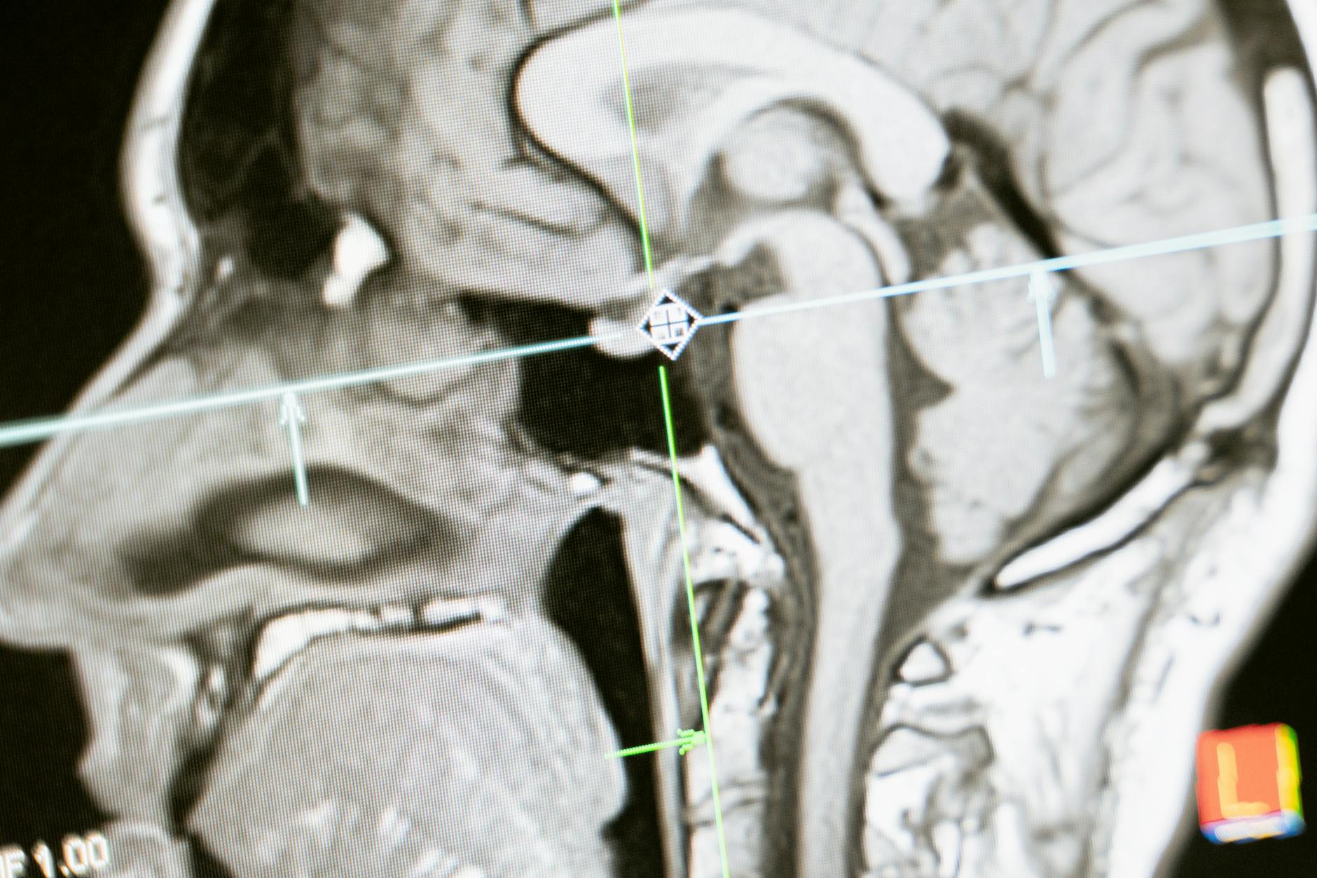
Radiologists identify encephalomalacia by its characteristic appearance across different MRI sequences. On T2 FLAIR imaging (a sequence sensitive to fluid), encephalomalacia typically shows as a dark signal surrounded by a bright rim of gliosis—scar tissue where glial cells have filled in the space left by dead neurons. This pattern is distinctive enough that experienced radiologists can usually recognize it immediately. The dark center represents cerebrospinal fluid that has replaced the lost brain tissue, while the bright rim represents the body’s attempt to wall off and stabilize the damaged area.
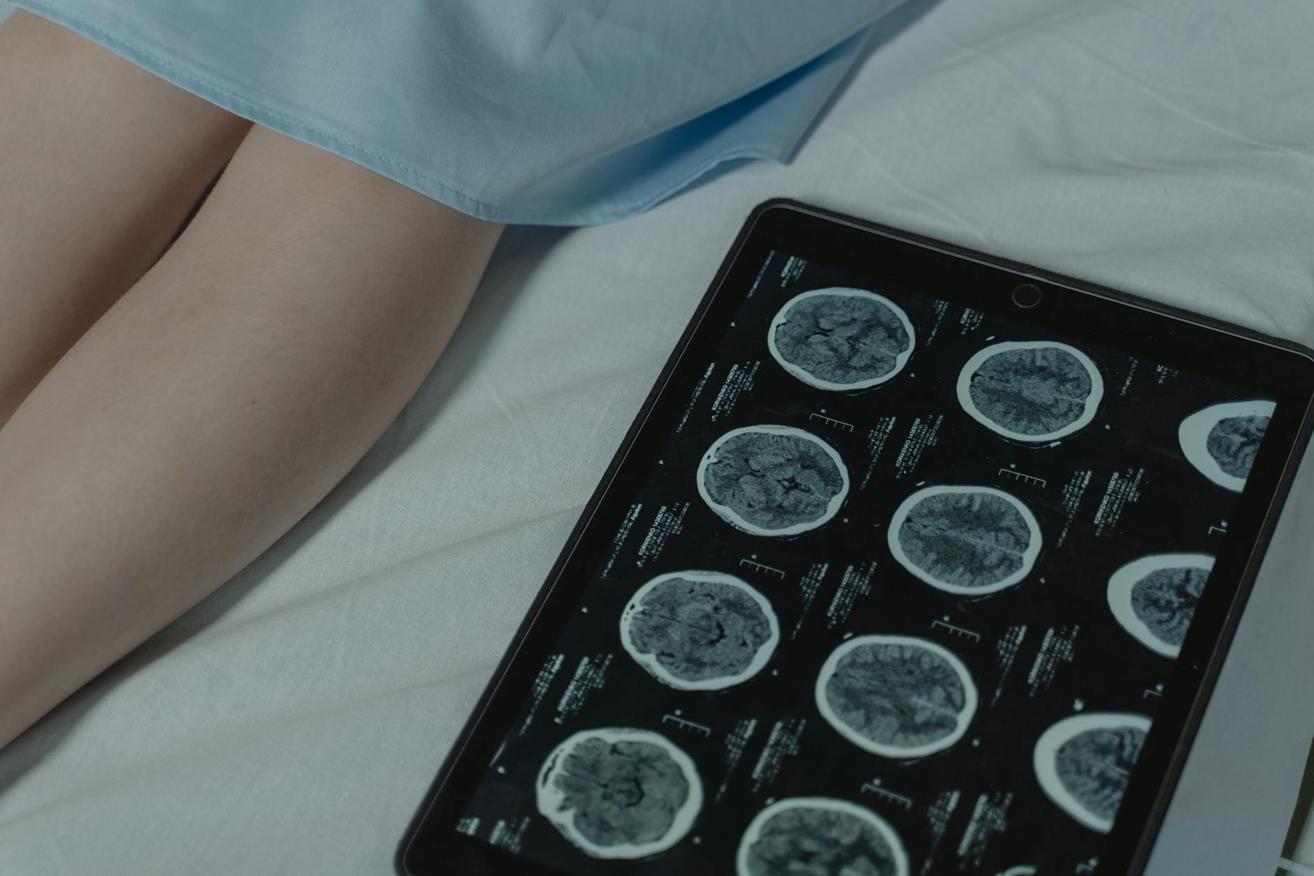
Understanding MRI sequences matters because different sequences highlight different aspects of brain pathology. On standard T2-weighted images, the fluid appears bright white, whereas on T1-weighted images, it appears dark. A radiologist interprets all these sequences together to confirm the diagnosis and assess whether the encephalomalacia is acute or chronic, improving or stable. In patients with dementia or cerebrovascular disease, MRI might reveal multiple small areas of encephalomalacia indicating repeated small strokes that the patient may not have consciously noticed—sometimes called silent infarcts.
What Causes Encephalomalacia to Develop?
Stroke is the most common cause of encephalomalacia, particularly in older adults and those with cardiovascular risk factors like hypertension or atrial fibrillation. When a blood clot blocks an artery supplying the brain, or when a blood vessel ruptures, the affected tissue dies within minutes. Months later, imaging shows the characteristic encephalomalacia in that territory. Traumatic brain injury, from falls, motor vehicle accidents, or assaults, also frequently leads to encephalomalacia in the regions where the impact damage occurred.
Infections can cause encephalomalacia, particularly severe cases of meningitis or encephalitis where inflammation damages large areas of tissue. Perinatal injury—damage occurring around the time of birth—represents a significant cause in pediatric patients, especially in premature infants whose immature brains are more vulnerable to hypoxic-ischemic events. Recent CDC data shows that influenza-associated encephalopathy in children contributes to serious neurological complications, with 13% of pediatric influenza deaths during the 2024-25 season through February involving severe neurological complications. Less commonly, chronic conditions like repeated seizures, progressive neurodegeneration, or chronic hypoxia can lead to diffuse encephalomalacia over time. The key point: encephalomalacia always represents a past injury or insult; it never develops without a preceding event that killed brain tissue.
Clinical Significance and Prognosis
The most important fact about encephalomalacia is that it is permanent. The brain cannot regrow lost tissue or regenerate dead neurons. However, this doesn’t mean the prognosis is uniformly poor—much depends on the location, extent of damage, and how soon the initial injury was treated. In acute stroke, prompt intervention with clot-busting medications or mechanical thrombectomy can minimize the area of tissue death, and therefore minimize the eventual encephalomalacia that appears months later. This emphasizes why rapid response to stroke symptoms—remembering the acronym FAST (Face drooping, Arm weakness, Speech difficulty, Time to call emergency services)—is so critical.
The functional impact of encephalomalacia depends heavily on location. Small encephalomalacia in the occipital lobe might affect vision, while similar-sized damage in the hippocampus could significantly impair memory formation. Some patients with encephalomalacia experience no noticeable symptoms if the damage is in an area with redundant function or if the brain has successfully reorganized other areas to compensate. Others experience substantial cognitive decline, movement problems, or personality changes. A warning for caregivers: new cognitive or neurological symptoms in a patient with known encephalomalacia might indicate new brain damage rather than progression of the existing encephalomalacia, necessitating prompt re-imaging and medical evaluation.

Encephalomalacia in Children and Age-Specific Considerations
Children’s brains respond differently to injury than adult brains, making pediatric encephalomalacia particularly concerning. Neonates and very young children are more prone to encephalomalacia from hypoxic-ischemic injury because their brains contain higher amounts of premyelinating oligodendrocytes and significantly more water content than mature brains. This biological vulnerability means that birth complications, respiratory distress, or infection poses a particular risk for developing encephalomalacia in the first weeks and months of life.
A clinical study of 50 children with cystic encephalomalacia admitted between January 2008 and December 2020 demonstrated that early-life hypoxic-ischemic events remain a major trigger for this condition in pediatric patients. The long-term implications can be severe: children with significant encephalomalacia from perinatal injury may face developmental delays, cerebral palsy, or intellectual disability depending on the extent and location of damage. However, the pediatric brain also demonstrates remarkable plasticity in many cases—younger children sometimes recover surprising amounts of function through intensive rehabilitation that leverages the brain’s neuroplastic capacity.
Management, Monitoring, and Future Directions
When encephalomalacia is discovered on imaging, the clinical focus shifts to managing symptoms, preventing new brain damage, and optimizing remaining function. This means addressing the underlying causes: controlling blood pressure and cholesterol to prevent future strokes, managing seizure disorders if they develop, treating infections, and participating in cognitive and physical rehabilitation. For patients with dementia, discovering encephalomalacia from previous strokes helps explain cognitive patterns and guides care planning.
Neuroimaging technology continues to advance. Newer MRI sequences and techniques can provide increasingly detailed information about brain tissue integrity and connectivity, potentially helping clinicians predict outcomes more accurately and tailor rehabilitation approaches. Research into neuroplasticity and brain reorganization offers hope that in the future, targeted interventions might enhance the brain’s ability to compensate for lost tissue. Currently, however, the emphasis remains on preventing further damage through aggressive management of vascular risk factors and maintaining cognitive and physical activity to support the brain’s adaptive capacity.
Conclusion
Encephalomalacia on a brain MRI indicates permanent loss of brain tissue where dead neurons have been replaced by cerebrospinal fluid—a finding that represents irreversible damage but not necessarily a dire prognosis. Understanding this distinction matters: while the brain cannot regrow lost tissue, the impact depends on location, extent, cause, and the individual’s age and overall brain health. The appearance of encephalomalacia signals that a past injury has occurred and healed in the way damaged brain tissue heals—by being reabsorbed and replaced with fluid.
If you or a loved one has received an encephalomalacia finding on an MRI, work with your healthcare team to identify the underlying cause, address modifiable risk factors, and develop a plan for managing symptoms and maintaining cognitive function. This might include stroke prevention measures, seizure management if needed, cognitive rehabilitation, and ongoing monitoring. The future of neurological care increasingly focuses on enhancing brain plasticity and supporting the nervous system’s remarkable ability to adapt—even when confronting permanent structural damage.
You Might Also Like
- What Do White Matter Changes on a Brain MRI Mean?
- Microangiopathic Changes on Brain MRI: What It Means
- Can Microangiopathic Changes Cause Memory Problems?
For more, see Alzheimer’s Association.





