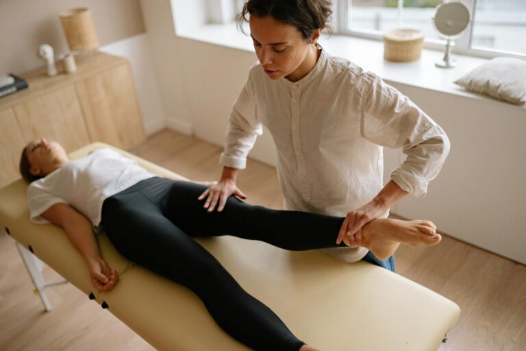
The sacroiliac joint (SI joint) is a frequently overlooked source of lower back and leg pain, affecting an estimated 15 to 30 percent of people with chronic, nonradicular pain. SI joint dysfunction develops when this joint—which connects the base of the spine to the pelvis—becomes unstable or inflamed, and certain risk factors significantly increase the likelihood of developing this condition. The nine primary risk factors are female sex, pregnancy, prior lumbar spine surgery, obesity, occupational or athletic overuse, leg length discrepancy, scoliosis, increased lumbar lordosis or anterior pelvic tilt, and age-related cartilaginous degeneration.
For example, a woman who undergoes lumbar fusion surgery and is overweight faces compounded risk from multiple factors simultaneously, making her particularly vulnerable to SI joint problems. Understanding these risk factors matters because SI joint dysfunction is often misdiagnosed as sciatica, herniated disc disease, or other spine conditions, leading to inappropriate treatment. About 25 percent of adults diagnosed with chronic low back pain actually have SI joint involvement as a primary or contributing factor. By recognizing your own risk profile, you can take steps to prevent symptoms or seek earlier intervention if pain develops.
Table of Contents
- Why Biological Sex and Pregnancy Create Higher Risk for SI Joint Dysfunction
- How Spinal Surgery and Structural Misalignment Drive SI Joint Problems
- The Weight and Overuse Connection in SI Joint Strain
- Biomechanical Imbalances That Destabilize the Sacroiliac Joint
- Age-Related Changes and the Two Peaks of SI Joint Dysfunction
- Understanding Prevalence Patterns and Who Is Most Affected
- Prevention and Management Implications
- Conclusion
Why Biological Sex and Pregnancy Create Higher Risk for SI Joint Dysfunction
Women are significantly more likely to develop SI joint dysfunction than men, a disparity rooted in biomechanics and hormonal physiology. The female pelvis is naturally wider and more mobile than the male pelvis, and the ligaments surrounding the SI joints are more lax—a protective adaptation for childbearing that paradoxically increases vulnerability to SI joint strain. This anatomical difference alone places women at higher baseline risk even without pregnancy. Pregnancy substantially amplifies this risk.
During pregnancy, the body releases relaxin, a hormone that increases ligament laxity throughout the pelvis to prepare for labor. This hormonal joint loosening, combined with the anterior weight shift created by a growing belly, places enormous stress on the SI joints. Pregnancy is identified in medical literature as one of the two most common risk factors for SI joint dysfunction, alongside prior lumbar spine surgery. Many women experience SI joint pain during pregnancy or within months after delivery; the ligaments typically restabilize gradually over weeks to months postpartum, though some women develop chronic dysfunction.

How Spinal Surgery and Structural Misalignment Drive SI Joint Problems
Prior lumbar fusion surgery ranks as the second most common risk factor for SI joint dysfunction, second only to pregnancy. When the lumbar spine is fused—typically to address disc herniation, stenosis, or instability—the segment above the fusion must compensate with increased motion, and the pelvis and SI joints absorb added stress. A patient who had lumbar fusion years earlier may develop SI joint pain well after the surgical site has healed and integrated. This is a biomechanical cascade effect rather than a surgical complication per se.
Beyond fusion surgery, structural spinal variations increase risk significantly. Scoliosis, an abnormal side-to-side curvature of the spine, creates uneven loading across the pelvis and predisposes the SI joints to asymmetric stress and micromotion. Similarly, increased lumbar lordosis—excessive inward curvature of the lower spine—and anterior pelvic tilt shift the mechanical advantage of the core muscles and place greater demand on the SI joint ligaments to maintain stability. However, having scoliosis or anterior pelvic tilt does not guarantee SI joint dysfunction; many people with these structural variations remain asymptomatic if they maintain adequate core strength and avoid aggravating movements.
The Weight and Overuse Connection in SI Joint Strain
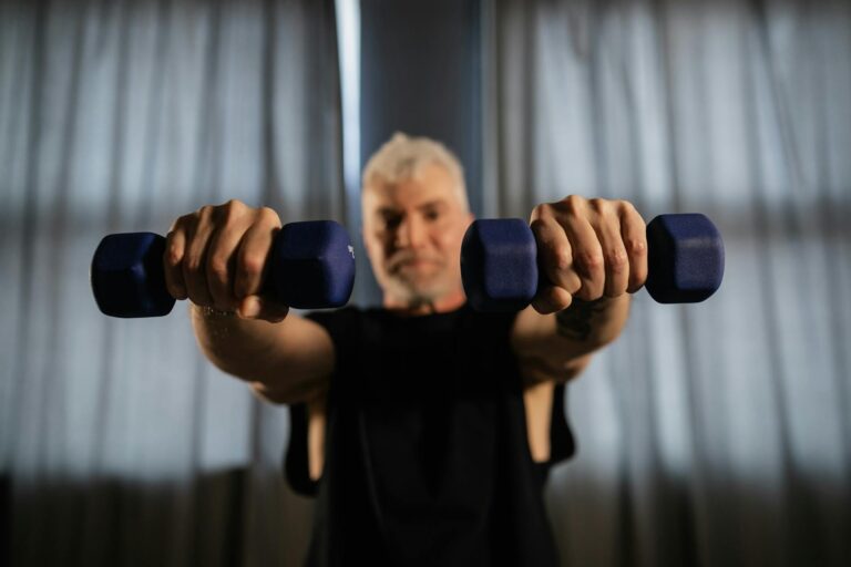
Obesity places cumulative mechanical stress on the SI joints and supporting ligaments. Excess body weight, particularly when concentrated in the abdomen, shifts the center of gravity forward, requiring the SI joints to work harder to stabilize the pelvis during standing, walking, and bending. Combined with reduced physical activity that often accompanies weight gain, weakened core muscles fail to provide adequate dynamic support, forcing the passive SI joint structures to bear more load than they are designed to handle.
Occupational and athletic overuse injuries represent another category of SI joint risk. Construction workers, nurses, and warehouse staff who lift heavy objects repeatedly without proper body mechanics, as well as runners and CrossFit athletes who perform high-impact movements, accumulate microtrauma in the SI joint ligaments. A specific example: a competitive distance runner who increases weekly mileage by more than 10 percent without allowing adequate recovery time can develop SI joint pain as the repetitive impact exceeds the joint’s adaptive capacity. Unlike sudden injuries, overuse-related SI joint dysfunction develops gradually and may not be noticed until pain becomes persistent.

Biomechanical Imbalances That Destabilize the Sacroiliac Joint
Leg length discrepancy—when one leg is structurally shorter than the other or when muscles are chronically shortened on one side—creates a functional leg length difference that tilts the pelvis. This tilting forces one SI joint into a compressed position while the other is pulled into a stretched, unstable position. Even a difference of one-half inch can alter pelvic mechanics significantly over time. Someone with a true leg length discrepancy of three-quarters of an inch might benefit from a shoe lift on the shorter side, yet many people with mild discrepancies resolve their SI pain through targeted stretching and strengthening rather than orthotics.
The relationship between pelvic tilt and SI joint health highlights the interdependence of spinal alignment and lower extremity mechanics. Anterior pelvic tilt—where the front of the pelvis drops forward and the tailbone tilts backward—flattens the lumbar curve and shifts muscular tension patterns. The gluteal muscles weaken while hip flexors tighten, creating a muscular imbalance that destabilizes the SI joints. Addressing this pattern requires not just stretching tight muscles but retraining postural habits and strengthening posterior chain muscles, a process that takes weeks of consistent effort.
Age-Related Changes and the Two Peaks of SI Joint Dysfunction
SI joint dysfunction follows a bimodal age distribution, meaning it occurs most frequently in two age groups: younger adults and older adults. In younger people, dysfunction typically stems from pregnancy, sports injuries, or occupational overuse. In adults over 50, age-related cartilaginous degeneration becomes the primary driver. The articular cartilage that lines the SI joint begins to thin in puberty and continues to degrade throughout life, eventually losing its ability to distribute forces evenly across the joint surface. This creates rougher joint surfaces, increased friction, and inflammation.
A common misconception is that SI joint pain in older adults is simply “normal aging” that cannot be treated. However, while age-related cartilage changes are inevitable, they do not automatically cause pain or dysfunction. Many people in their 60s, 70s, and 80s have asymptomatic SI joint changes visible on imaging. Pain develops when degenerative changes are combined with reduced core strength, decreased flexibility, and altered movement patterns. Maintaining strength and mobility throughout life, therefore, is one of the few modifiable factors that can delay or prevent symptomatic SI joint degeneration.

Understanding Prevalence Patterns and Who Is Most Affected
Approximately 25 percent of people with chronic low back pain have SI joint involvement, yet this diagnosis is often missed because SI pain can masquerade as typical lower back pain, sciatica, or hip pain. Among individuals with chronic, nonradicular pain—pain that does not follow a specific nerve distribution—the prevalence rises to 15 to 30 percent, making SI joint dysfunction one of the more common but underrecognized pain generators. The bimodal age distribution reflects these underlying risk patterns clearly.
A 35-year-old postpartum woman and a 65-year-old retired laborer with degenerative changes are both at high risk, but for different reasons. The younger woman’s risk is time-limited; her ligaments will restabilize over months to a couple of years if she manages activity appropriately. The older man’s risk is more persistent because cartilage does not regenerate, though appropriate exercise can often prevent pain even with degenerative changes present.
Prevention and Management Implications
The presence of risk factors does not mean SI joint dysfunction is inevitable. Someone with multiple risk factors—for instance, a middle-aged woman who is overweight and has a history of lumbar fusion—can still prevent SI pain through targeted prevention strategies: maintaining a healthy weight, strengthening core and gluteal muscles, correcting postural imbalances, and using proper body mechanics during lifting. These modifications are within individual control.
Forward-looking, research continues to clarify the best interventions for SI joint dysfunction at different stages of the lifecycle. For pregnant women, specialized SI joint belts during pregnancy and early postpartum can reduce pain and improve function. For older adults, exercise programs that combine strength and stability training show promise in managing degenerative changes. Understanding your personal risk profile allows you to tailor prevention efforts to the factors most relevant to your situation.
Conclusion
SI joint dysfunction is a highly prevalent condition affecting millions of adults, yet it remains underdiagnosed because its symptoms often overlap with other lower back pain syndromes. The nine primary risk factors—female sex, pregnancy, prior lumbar fusion, obesity, occupational or athletic overuse, leg length discrepancy, scoliosis, increased lumbar lordosis, and age-related degeneration—operate through biomechanical and structural pathways that can be understood and, in many cases, modified.
A woman’s hormonal changes, a manual laborer’s repetitive stress, a surgeon’s fused spine, or a runner’s training error all represent distinct but addressable risk pathways. If you experience lower back pain, buttock pain, or hip pain—particularly pain on one side—and standard low back pain treatments have not worked, consider asking your healthcare provider about SI joint dysfunction. Recognizing your risk factors and addressing modifiable ones through weight management, targeted exercise, proper body mechanics, and postural correction can often prevent symptoms or significantly reduce their severity.