When doctors talk about brain reserve and vascular burden, they’re describing two separate but interconnected biological systems that determine how well your brain resists cognitive decline. Brain reserve refers to the structural surplus your brain accumulated over a lifetime—the extra neurons, synapses, and brain tissue that act as a buffer against aging and disease. Vascular burden, by contrast, measures the damage to blood vessels in the brain from chronic conditions like high blood pressure, diabetes, and smoking, visible on brain imaging as white matter hyperintensities (WMH). Together, these concepts help explain why two people with similar levels of Alzheimer’s pathology can experience vastly different cognitive outcomes: one person’s substantial brain reserve protects them longer, while another’s high vascular burden accelerates their decline.
Think of brain reserve as your neural savings account. A 75-year-old with decades of education, mentally stimulating work, and rich social connections accumulated more synaptic density and larger gray matter volumes than a 75-year-old without those experiences. When age-related neural loss begins—which it does in everyone—the person with greater reserve can tolerate more loss before cognitive impairment becomes noticeable. Vascular burden, meanwhile, directly undermines this protection by starving brain tissue of oxygen and nutrients, creating a race between reserve and damage.
Table of Contents
- How Do Doctors Measure Brain Reserve in Living Patients?
- What Are White Matter Hyperintensities and Why They Matter
- How Brain Reserve and Vascular Burden Interact
- Why Doctors Now Take Vascular Burden Seriously for Dementia Risk
- What Makes Brain Reserve Difficult to Quantify in Current Practice
- How Lifespan Factors Shape Brain Reserve and Vascular Burden
- Vascular Burden Can Progress Silently Until Cognitive Symptoms Appear
- Frequently Asked Questions
How Do Doctors Measure Brain Reserve in Living Patients?
Brain reserve isn’t a single number that doctors can check with a blood test. Instead, it’s inferred from multiple indicators: structural brain imaging showing total brain volume and cortical thickness, cognitive testing documenting baseline mental abilities, and detailed life history covering education, occupation, cognitive activities, and social engagement. Neuropsychologists estimate reserve by comparing a patient’s current cognitive performance against their expected baseline—someone who was a surgeon or professor and is now showing early memory loss is presumed to have started from a higher baseline than someone without that background.
The challenge is that brain reserve exists on a spectrum with no standardized, validated measurement across research institutions. A person with a PhD and 40 years in intellectual work has more reserve than a person with a high school diploma and manual labor experience, but quantifying that difference remains imprecise. This matters clinically: without knowing someone’s premorbid reserve, doctors can’t reliably predict when their cognitive decline will become noticeable, or whether mild test findings represent early disease or simply normal aging within the bounds of a smaller starting reserve.
What Are White Matter Hyperintensities and Why They Matter
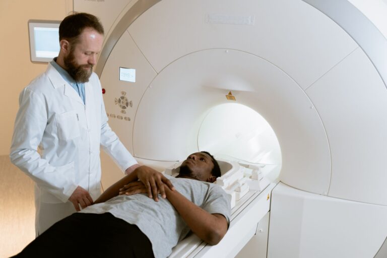
White matter hyperintensities are bright areas visible on MRI scans, appearing as spots or streaks in the brain’s white matter—the tissue containing nerve fibers that connect different brain regions. They represent areas of chronic ischemia (reduced blood flow) and hypoxia (insufficient oxygen), typically caused by vascular risk factors: uncontrolled hypertension is the strongest driver, followed by diabetes, obesity, smoking history, and previous stroke. Research quantifies vascular burden using a Vascular Burden Index that combines the volume of white matter hyperintensities with other imaging markers of cerebrovascular disease.
The cognitive impact is measurable and specific. Studies using brain imaging and cognitive testing show that larger white matter hyperintensity volumes correlate with worse performance across all cognitive domains—memory, processing speed, language, and executive function—with the strongest effect on semantic fluency (retrieving category words like “animals” or “tools” under time pressure). A patient with extensive white matter hyperintensities might score within normal range on standard cognitive screening but show obvious difficulty when pushed, and their cognitive decline typically accelerates compared to someone with minimal vascular burden. Critically, this damage is independent of Alzheimer’s pathology—someone without amyloid or tau can still develop cognitive impairment from vascular burden alone.
How Brain Reserve and Vascular Burden Interact
Brain reserve and vascular burden work against each other. A person with high reserve and minimal vascular burden can maintain normal cognition well into advanced age despite accumulating some neuropathology. Someone with low reserve and high vascular burden risks earlier, faster cognitive decline. But the interaction is not simply additive—when both are present simultaneously, the effect accelerates.
Research shows that patients with both Alzheimer’s disease AND cerebrovascular disease (high vascular burden) show markedly faster cognitive deterioration compared to people with either condition alone, suggesting that vascular damage actively undermines the brain’s ability to compensate. A concrete example: two 80-year-old women both have similar amyloid-tau pathology on biomarker testing, but one maintained a career as a teacher, stayed socially active, learned new skills (higher brain reserve), and has minimal white matter hyperintensities on MRI. The other had less formal education, fewer cognitively demanding activities, and extensive white matter hyperintensities from decades of uncontrolled hypertension. The first woman shows normal memory and language; the second shows mild cognitive impairment affecting multiple domains. Their Alzheimer’s pathology is comparable, but their clinical presentation differs dramatically because of reserve and vascular burden.
Why Doctors Now Take Vascular Burden Seriously for Dementia Risk
For decades, dementia research focused almost exclusively on Alzheimer’s pathology—amyloid and tau plaques in the brain. Vascular contributions to cognitive decline were considered secondary or even irrelevant in Alzheimer’s disease. That view has shifted substantially. Large autopsy studies show that the majority of cognitively impaired older adults have mixed pathology—Alzheimer’s changes AND cerebrovascular disease—rather than pure Alzheimer’s disease.
Large population studies document that vascular burden is independently associated with higher dementia risk and faster cognitive decline, even after accounting for Alzheimer’s biomarkers. This shift matters practically because vascular burden is modifiable, whereas Alzheimer’s pathology remains difficult to prevent or reverse. Blood pressure control, diabetes management, smoking cessation, weight management, and treatment of cardiovascular disease all reduce vascular burden accumulation and slow cognitive decline. Epidemiologic data from high-income countries show declining dementia rates over recent decades, partly attributed to better cardiovascular risk management—fewer people with severe uncontrolled hypertension, better diabetes control, and reduced smoking rates. By contrast, countries without widespread cardiovascular management show rising dementia prevalence, suggesting that controlling vascular risk factors genuinely prevents or delays cognitive impairment.
What Makes Brain Reserve Difficult to Quantify in Current Practice
A significant limitation in dementia research and clinical practice is the lack of standardized, validated methods for measuring or quantifying brain reserve. Different studies use different proxy measures: years of education, occupational complexity, scores from cognitive training questionnaires, structural brain volume, or combinations of these. A person with 16 years of formal education but minimal cognitive engagement in adulthood may have lower reserve than a high school graduate who spent 40 years solving complex problems at work. The correlation between education and reserve is moderate, not perfect.
Another limitation: reserve is dynamic and can decline through disuse. Someone who was highly educated but stops engaging cognitively—no reading, no novel learning, social withdrawal—may see their reserve atrophy somewhat, though probably not to the extent they would have had if they never built it. This creates uncertainty when assessing dementia risk in older adults: doctors can estimate premorbid reserve from life history, but current reserve depends on ongoing cognitive and social engagement, which varies widely and changes over time. This uncertainty also means that even sophisticated dementia risk models still have limited ability to predict individual outcomes—population trends are clear, but person-by-person prediction remains imprecise.
How Lifespan Factors Shape Brain Reserve and Vascular Burden
Brain reserve doesn’t develop equally for everyone. Education, occupation, cognitive hobbies, social engagement, physical fitness, and absence of early-life adversity all contribute to reserve building. But research increasingly shows that social determinants of health—income, neighborhood environment, racial and ethnic discrimination, access to quality education and healthcare—profoundly shape reserve accumulation and vascular risk exposure. A person growing up in poverty, experiencing chronic stress, with limited educational access, and in a neighborhood with poor healthcare access both builds less reserve and accumulates vascular risk factors earlier in life.
Early-life adversity also leaves direct marks on reserve capacity. Childhood maltreatment, severe trauma, or chronic stress in formative years associates with smaller brain volumes and reduced synaptic density in adulthood, suggesting reserve capacity is constrained from the start. This doesn’t mean early adversity guarantees cognitive impairment later, but it means that all else being equal, two people with equal educational attainment may start adulthood with different reserve ceilings based on their early environments. This highlights why dementia prevention isn’t purely individual—it’s rooted in social structures and inequalities that accumulate across the lifespan.
Vascular Burden Can Progress Silently Until Cognitive Symptoms Appear
One of the most clinically important aspects of vascular burden is that white matter hyperintensities accumulate silently, without subjective symptoms, over years or decades. A person with 20 years of poorly controlled hypertension may have extensive white matter hyperintensities on brain MRI but report no cognitive complaints and score normally on bedside cognitive screening. The damage is happening—neurons are being hypoxic, synapses are being lost, white matter connectivity is degrading—but cognitive reserve is still compensating, masking the decline. Then, at some point, reserve becomes exhausted relative to the accumulated damage, and cognitive symptoms emerge abruptly or accelerate.
This is why vascular risk factor management in midlife and early older adulthood is crucial for dementia prevention, even though people feel cognitively fine. A 55-year-old with blood pressure consistently 150/90 is accumulating white matter disease silently. A 65-year-old smoker with diabetes is experiencing ongoing hypoxic stress in their brain. By the time cognitive decline becomes clinically apparent at age 75 or 80, the damage is extensive and difficult to reverse with medication alone. The evidence supports aggressive control of hypertension, diabetes, cholesterol, and smoking starting in middle age, decades before any cognitive symptoms emerge, as the most effective way to reduce vascular burden’s impact on dementia risk.
Frequently Asked Questions
Can you increase your brain reserve after age 60?
Yes, but probably not as dramatically as earlier in life. Cognitive engagement, learning new skills, social connection, and physical exercise still build or maintain reserve through neuroplasticity in older adults. However, the brain’s structural capacity to add new neurons and synapses declines with age, so the gains are more modest than what education and complex work build in younger people.
Are white matter hyperintensities the same as stroke?
No. White matter hyperintensities represent chronic ischemia and hypoxia from vascular disease, visible on MRI as damage zones. A stroke is an acute event—sudden blockage or rupture of a brain blood vessel—causing rapid neuronal death in a localized area. However, chronic white matter disease increases stroke risk, and small accumulating silent strokes can contribute to white matter hyperintensity burden.
Can blood pressure medication reduce white matter hyperintensities that already exist?
Studies show that aggressive blood pressure control can slow the progression of white matter hyperintensities and prevent new ones from forming, but reversing existing extensive white matter damage is difficult. This underscores why prevention—keeping blood pressure well-controlled from midlife onward—is more effective than trying to reverse damage years later.
Does cognitive training increase brain reserve the same way education does?
Cognitive training and learning new complex skills can maintain or modestly enhance reserve, but the effect is typically smaller and more narrow than years of formal education and intellectually demanding work. Training tends to improve performance on the trained task itself (like crossword puzzles or memory games) but may not generalize to broad cognitive protection against aging and disease.
If I have high vascular burden, can excellent brain reserve prevent dementia?
Substantial brain reserve can substantially delay or reduce dementia symptom onset despite vascular burden, but cannot eliminate the risk entirely. It’s protective but not protective to the point of immunity. This is why managing both vascular risk factors (to reduce burden) and engaging in reserve-building activities (education, cognitive engagement, social connection) matters—both reduce dementia risk.
What imaging test shows vascular burden most clearly?
MRI is the standard. It visualizes white matter hyperintensities, which are the primary marker of vascular burden in clinical and research settings. A brain MRI in an older adult with cognitive concerns will typically include specific sequences that highlight white matter changes. CT scans can detect some severe vascular changes but are less sensitive than MRI for detecting white matter disease.