Types of Diagnostic Scans for Screening Dementia and Alzheimer’s – CT, MRI, PET Scans

There are several types of scans that may be used to help diagnose and monitor dementia, including:
- CT scans: CT scans use X-rays to create detailed images of the brain’s structure. They can help in detecting structural changes in the brain associated with dementia, such as atrophy (shrinking) of the brain, enlarged ventricles (fluid-filled spaces in the brain), or the presence of brain lesions.
- MRI scans: MRI scans use a magnetic field and radio waves to create detailed images of the brain’s structure. They can provide more detailed images of soft tissues, such as the brain, spinal cord, and internal organs, and can be used to detect changes in the brain associated with dementia.
- PET scans: PET scans use a small amount of radioactive material to create images of the brain’s metabolic activity. They can detect the presence of amyloid plaques and tau tangles in the brain, which are the hallmark features of Alzheimer’s disease.
- SPECT scans: SPECT (Single Photon Emission Computed Tomography) scans are similar to PET scans but use a different type of radioactive material. They can be used to detect changes in blood flow in the brain, which can be useful in diagnosing certain types of dementia.
- Cerebrospinal fluid analysis: Cerebrospinal fluid (CSF) is the fluid that surrounds the brain and spinal cord. Analysis of CSF can provide valuable information about the presence of certain proteins or biomarkers that are associated with different types of dementia, including Alzheimer’s disease.
The choice of imaging technique for detecting other types of dementia will depend on the specific type of dementia and the individual patient’s symptoms and medical history.
For example, in vascular dementia, which is caused by reduced blood flow to the brain, MRI and CT scans can be useful in detecting changes in the brain’s blood vessels, such as strokes or other abnormalities. PET scans can also be used to evaluate blood flow in the brain.
In frontotemporal dementia, which affects the front and side areas of the brain, MRI and PET scans can help in detecting changes in these areas of the brain, such as atrophy or shrinkage.
In Lewy body dementia, which is caused by abnormal protein deposits in the brain, imaging techniques such as SPECT (Single Photon Emission Computed Tomography) and PET scans can be used to detect the presence of these protein deposits in the brain.
Both CT scans and MRIs can help in detecting neurological disorders such as dementia, although they are not typically used as standalone diagnostic tests for this condition.
A CT scan can detect changes in brain tissue that may indicate the presence of dementia, such as atrophy (shrinking) of the brain, enlarged ventricles (fluid-filled spaces in the brain), or the presence of brain lesions.
An MRI scan can also help in detecting structural changes in the brain associated with dementia, such as shrinkage of the hippocampus (a region of the brain involved in memory formation and recall). Additionally, MRI can detect changes in brain chemistry and blood flow that may be associated with dementia.
However, dementia is a complex condition that often requires a combination of diagnostic tests, including medical history, neurological exams, cognitive tests, and imaging tests, to make a diagnosis. Therefore, it is important to consult with a healthcare professional for an accurate diagnosis and appropriate treatment.
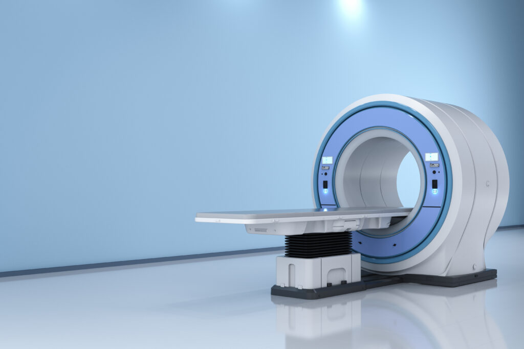
An MRI (Magnetic Resonance Imaging) is a medical imaging technique that uses a powerful magnetic field and radio waves to generate detailed images of the internal structures of the body. Unlike CT scans, MRI scans do not use ionizing radiation.
During an MRI, the patient lies inside a large tube-like machine, and the magnetic field and radio waves cause the hydrogen atoms in the body’s cells to produce a signal that is detected by the machine. This signal is then used to create high-resolution images of the body’s tissues and organs.
MRI scans can provide more detailed images of soft tissues, such as the brain, spinal cord, and internal organs, and can be used to diagnose and monitor a wide range of medical conditions, including cancer, neurological disorders, and musculoskeletal injuries. MRI is also used to evaluate the health of organs such as the liver and heart.
Both CT scans and MRIs can help in detecting neurological disorders such as dementia, although they are not typically used as standalone diagnostic tests for this condition.
A CT scan can detect changes in brain tissue that may indicate the presence of dementia, such as atrophy (shrinking) of the brain, enlarged ventricles (fluid-filled spaces in the brain), or the presence of brain lesions.
An MRI scan can also help in detecting structural changes in the brain associated with dementia, such as shrinkage of the hippocampus (a region of the brain involved in memory formation and recall). Additionally, MRI can detect changes in brain chemistry and blood flow that may be associated with dementia.
However, dementia is a complex condition that often requires a combination of diagnostic tests, including medical history, neurological exams, cognitive tests, and imaging tests, to make a diagnosis. Therefore, it is important to consult with a healthcare professional for an accurate diagnosis and appropriate treatment.
CT scans can help in detecting structural changes in the brain associated with Alzheimer’s disease, but they are not typically used as a standalone diagnostic tool for this condition.
During the early stages of Alzheimer’s disease, CT scans may show changes in the brain structure, such as shrinkage of the hippocampus (a region of the brain involved in memory formation and recall), but these changes can also be observed in the aging brain and in other conditions, such as vascular dementia. Additionally, CT scans cannot detect the specific brain abnormalities, such as amyloid plaques and tau tangles, that are characteristic of Alzheimer’s disease.
In most cases, a diagnosis of Alzheimer’s disease requires a combination of diagnostic tests, including medical history, neurological exams, cognitive tests, and imaging tests such as MRI, PET scans, or cerebral spinal fluid analysis. Therefore, it is important to consult with a healthcare professional for an accurate diagnosis and appropriate treatment.
What About PET Scans?
A PET (Positron Emission Tomography) scan is a medical imaging technique that uses a small amount of radioactive material (known as a radiotracer) to create images of the body’s metabolic activity.
During a PET scan, the patient is injected with a radiotracer, which is then absorbed by the body’s cells. The radiotracer releases particles called positrons, which collide with electrons in the body’s cells and produce gamma rays. These gamma rays are detected by a special camera that creates a 3D image of the body’s metabolic activity.
PET scans can be used to diagnose and monitor a wide range of medical conditions, including cancer, neurological disorders, and cardiovascular disease. In Alzheimer’s disease, PET scans can detect the presence of amyloid plaques and tau tangles, which are characteristic of the disease. PET scans can also help in evaluating the effectiveness of treatment for certain conditions and in planning surgeries or radiation therapy.
Which Scans are the Best at Diagnosing Alzheimer’s Disease?
Though, this is may be a controversial question, out of CT scans, MRIs, and PET scans, PET scans are considered the most effective at detecting Alzheimer’s disease, particularly in the early stages. PET scans can detect the presence of amyloid plaques and tau tangles in the brain, which are the hallmark features of Alzheimer’s disease. However, PET scans are typically more expensive and involve exposure to a small amount of radiation. MRIs can also provide valuable information in detecting Alzheimer’s disease by detecting structural changes in the brain, such as shrinkage of the hippocampus, but are not as effective at detecting the specific biomarkers of the disease as PET scans. CT scans are less sensitive than both MRI and PET scans in detecting Alzheimer’s disease and are not typically used as a primary diagnostic tool for the condition. Ultimately, the choice of imaging technique will depend on a variety of factors, including the patient’s medical history, symptoms, and the preferences of the healthcare provider.