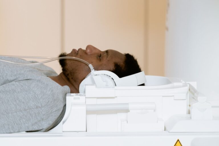
A head CT scan fits into an Alzheimer's workup as one supporting test, not a diagnosis by itself. Its central job is to rule out treatable causes of memory and thinking problems. Alzheimer's diagnosis combines medical history, neurological exams, cognitive and functional assessments, brain imaging, and spinal-fluid or blood tests, according to the Alzheimer's Association medical tests guide. A CT result therefore guides next steps rather than ends the evaluation.
Medical information disclaimer: This article is for general educational purposes only and does not provide medical advice, diagnosis, or treatment. Always consult a physician or other qualified health professional about symptoms, medications, tests, or treatment decisions.
Table of Contents
- What a head CT rules out
- Why the usual order is CT without contrast
- Why CT cannot confirm Alzheimer's alone
- What usually comes after the CT
What a head CT rules out
In adults evaluated for dementia or mild cognitive impairment, CT plays an exclusion role. It helps rule out tumor, stroke, bleeding, and fluid buildup that can mimic dementia, according to RadiologyInfo dementia imaging guide.
Finding one of these causes can change treatment quickly. The same scan can also show brain shrinkage linked to dementia. It gives detailed information about head injury, stroke, and other brain diseases that explain cognitive symptoms.
Why the usual order is CT without contrast
For initial imaging of mild memory problems and typical suspected Alzheimer disease, noncontrast head CT is rated "Usually Appropriate" by the American College of Radiology dementia imaging guidance. That rating makes it a common first structural screen. It is fast and widely available.
CT with contrast, or CT without-and-with contrast, is rated "Usually Not Appropriate" for that first Alzheimer-type workup. So the standard CT order is noncontrast. Contrast adds little for this question and adds risk and complexity.
Why CT cannot confirm Alzheimer's alone
CT alone cannot definitively diagnose Alzheimer's disease. Its detail is limited for early brain changes. MRI is generally preferred when available because it shows finer brain detail without ionizing radiation.
The radiation difference is concrete but small for one scan. Head CT carries low-dose exposure, while MRI uses no ionizing radiation. Availability, speed, implants, kidney issues, and need for sedation can still make CT the practical first step.
What usually comes after the CT
A normal CT does not prove Alzheimer's. An abnormal CT may point to another cause.
Either way, doctors use more specific tests to confirm Alzheimer pathology. Those follow-up tests often include MRI and amyloid or tau PET, spinal-fluid testing, or blood biomarker testing. The sequence is practical and staged:.
- use noncontrast CT as the initial structural screen
- use MRI for finer structural detail when needed
- use PET, spinal-fluid, or blood tests to check Alzheimer markers