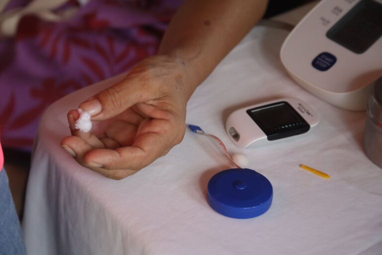
Doctors diagnose dementia through a series of cognitive tests, brain imaging scans, and blood work designed to identify memory loss and thinking changes while ruling out treatable conditions that mimic dementia symptoms. There is no single test that confirms dementia. Instead, a neurologist or geriatrician builds a diagnosis by combining cognitive assessments (which measure memory, language, and processing speed), structural imaging like MRI or CT scans (which show brain atrophy or other physical changes), and sometimes specialized blood tests that detect protein markers in the bloodstream.
A 68-year-old woman who started forgetting recent conversations might undergo an MRI to rule out a small stroke, a cognitive test to measure her memory decline, and blood work to exclude vitamin B12 deficiency—all before a doctor can say with confidence that she has Alzheimer’s disease. The diagnostic process reflects an important reality: dementia is a clinical diagnosis confirmed by pattern recognition, not by a single abnormal result. Doctors must rule out reversible causes—thyroid problems, depression, sleep apnea, medication interactions—that can produce dementia-like symptoms. This is why the evaluation process can take weeks or months, involves multiple doctor visits, and may require coordination between primary care physicians, neurologists, and sometimes psychiatrists or speech pathologists.
Table of Contents
- What Tests Are Actually Used to Diagnose Dementia?
- Brain Imaging: Seeing the Physical Evidence of Dementia
- Blood Tests and Biomarkers: The New Frontier in Dementia Diagnosis
- Ruling Out the Imposters: Conditions That Masquerade as Dementia
- The Lumbar Puncture: Cerebrospinal Fluid Testing When It’s Needed
- What to Expect During a Dementia Diagnostic Evaluation
- The Role of Specialist Consultation in Complex Cases
What Tests Are Actually Used to Diagnose Dementia?
The foundation of dementia diagnosis rests on cognitive screening tools administered in the office. The Montreal Cognitive Assessment (MoCA) is one of the most common; it takes 10 to 15 minutes and tests memory, language, visuospatial skills, and executive function through tasks like copying a clock face or recalling a short list of words. The Mini-Cog is even quicker—three minutes—and includes clock drawing and a three-word recall test; it’s designed for primary care settings where time is limited. If a screening tool suggests decline, a neuropsychologist may conduct more extensive testing over several hours, administering dozens of subtests to create a detailed cognitive profile.
This deeper evaluation distinguishes between normal aging (forgetting where you put your keys) and pathological decline (not remembering you own a car). Cognitive tests must be interpreted in context. A person with eight years of formal education may score lower on certain language tasks than a college graduate, not because of dementia but because of baseline differences in education and verbal experience. Some tests are sensitive to depression, which can mask itself as memory loss; a patient who is depressed and withdrawn may perform poorly on cognitive testing but recover normal function after antidepressant treatment. The limitation is this: cognitive tests measure current function but don’t tell you what’s causing the decline.
Brain Imaging: Seeing the Physical Evidence of Dementia
MRI (magnetic resonance imaging) and CT (computed tomography) scans show the brain’s structure and can reveal whether the ventricles are enlarged, whether there is brain atrophy, and whether small strokes have occurred silently in the brain. In Alzheimer’s disease, the hippocampus—a seahorse-shaped structure critical for memory—often shrinks visibly on MRI. In vascular dementia, white matter lesions (areas of reduced blood flow) appear as bright spots on certain MRI sequences. A PET (positron emission tomography) scan can show metabolic activity and the buildup of amyloid-beta and tau proteins, the hallmark proteins of Alzheimer’s disease, but PET is expensive and not available everywhere.
The catch is that brain imaging is not always definitive. Many cognitively normal 70-year-olds have small strokes on MRI or some degree of brain atrophy; asymptomatic amyloid buildup on PET imaging can precede symptoms by years or decades. Conversely, a patient with clear clinical dementia may have an MRI that looks relatively normal, especially early in disease. Imaging rules out stroke, tumor, and hydrocephalus (fluid buildup), but it doesn’t confirm the cause of dementia—it only rules out certain mimics and provides supporting evidence.
Blood Tests and Biomarkers: The New Frontier in Dementia Diagnosis
Blood biomarkers have transformed dementia diagnosis in the past few years. tests can now measure phosphorylated tau, amyloid-beta-42, and phosphorylated tau-181 directly from blood serum, offering a window into the pathological processes happening in the brain. These tests can show Alzheimer’s-type changes before symptoms appear and can predict who will decline cognitively in the coming years. A 62-year-old with memory complaints and a blood test showing elevated p-tau-181 and low amyloid-beta-42 has clear biological evidence of Alzheimer’s pathology. Biomarker testing also helps identify which dementia type someone is at risk for, informing treatment choices.
However, biomarker positivity is not the same as dementia. A cognitively normal person can have Alzheimer’s biomarkers and never develop dementia symptoms during their lifetime. The tests are also not uniformly available and can be expensive if not covered by insurance. Different laboratories use different cutoff values, which can lead to inconsistency in results. Biomarker tests are most useful when combined with cognitive testing and clinical judgment, not as standalone diagnostic tools.
Ruling Out the Imposters: Conditions That Masquerade as Dementia
A comprehensive dementia evaluation always includes blood tests to rule out thyroid disease, vitamin B12 deficiency, folate deficiency, syphilis, and other metabolic problems that can mimic cognitive decline. Depression is one of the most common masqueraders; older adults with major depression can present with memory problems, poor concentration, and social withdrawal that look identical to dementia. Testing for depression involves a depression screening questionnaire like the Geriatric Depression Scale and sometimes an interview by a psychiatrist. If depression is the culprit, antidepressant medication may restore cognitive function within weeks.
Sleep apnea causes daytime grogginess, poor memory consolidation, and mood changes that can be mistaken for early dementia. A sleep study (polysomnography) reveals whether a patient stops breathing repeatedly during sleep; treating sleep apnea often improves cognition. Medication interactions are another hidden culprit: anticholinergic drugs (used for overactive bladder or anxiety), benzodiazepines, and opioids can all impair memory and thinking. A careful medication review by a pharmacist or neurologist sometimes reveals that a patient’s “dementia” is actually drug-induced and reversible. The limitation here is that ruling out these conditions takes time—blood work must be ordered, results returned, sleep studies scheduled—which is why a complete dementia workup often takes months rather than weeks.
The Lumbar Puncture: Cerebrospinal Fluid Testing When It’s Needed
In some cases, a neurologist will recommend a lumbar puncture (spinal tap) to measure cerebrospinal fluid (CSF) biomarkers directly. CSF analysis can show tau tangles and amyloid-beta accumulation with high accuracy and can sometimes reveal other conditions like encephalitis or meningitis that cause dementia-like symptoms. A 55-year-old with rapidly progressive cognitive decline, psychiatric symptoms, and behavioral changes might undergo lumbar puncture to rule out infectious causes or prion diseases like Creutzfeldt-Jakob disease.
The barrier to lumbar puncture is patient acceptance and access. Many people fear the spinal tap, worry about headaches afterward, or have anatomical issues (severe osteoporosis, previous back surgery) that make the procedure difficult. Not all neurology practices perform lumbar punctures routinely, so a patient may have to travel to a specialized center. Most dementia diagnoses do not require lumbar puncture—it’s reserved for atypical presentations, rapid decline, or young patients where an unusual cause is suspected.
What to Expect During a Dementia Diagnostic Evaluation
A typical dementia evaluation begins with a detailed history from the patient and often a close family member or caregiver who can describe the pattern of cognitive changes over months or years. The primary care doctor or neurologist performs a physical and neurological exam, looking for evidence of stroke, Parkinson’s disease, or other conditions that might explain symptoms. Office-based cognitive screening tests follow—a 10-minute MoCA or Mini-Cog that serves as the initial filter. If results are abnormal, the patient is referred to a neurologist or neuropsychologist and scheduled for imaging. Blood work is ordered at the same time. If available and indicated, biomarker testing is discussed.
A follow-up appointment is scheduled to review all results together and form a diagnostic impression. This process places demands on patients and families. Multiple appointments are required; tests must be scheduled in sequence rather than simultaneously (imaging must happen before the follow-up visit to discuss findings). Patients may need time off work; caregivers may need to accompany them. Insurance may require pre-approval for certain tests, introducing delays. The entire evaluation from first symptom report to confirmed diagnosis often takes three to six months.
The Role of Specialist Consultation in Complex Cases
When cognitive decline is atypical, rapid, or includes unusual features like prominent behavioral changes or movement disorder, neurologists often refer patients to specialized memory clinics or cognitive neurology centers. These clinics may have neuropsychologists on staff who conduct extended testing; access to advanced imaging; and expertise in rare dementias like primary progressive aphasia, behavioral variant frontotemporal dementia, or Lewy body dementia. A patient presenting with apathy and loss of inhibition rather than memory loss might be suspected of frontotemporal dementia; a specialist can recognize this pattern and order the appropriate tests.
A person with memory loss plus tremor and rigid movements might have Lewy body disease, which requires different evaluation and treatment than Alzheimer’s disease. Access to specialist evaluation depends partly on geography and insurance. Rural patients may live hours from the nearest memory clinic; some insurance plans limit neurology referrals or require prior authorization. Patients diagnosed in a community setting without specialist input may receive a less specific diagnosis—”probable Alzheimer’s disease” rather than a confirmed type—which can affect access to clinical trials and emerging treatments.
- —