Reviewed by the Help Dementia Editorial Team — our editors review every article for accuracy against guidance from the National Institute on Aging, the Alzheimer’s Association, and peer-reviewed sources.
Yes, you can absolutely have dementia with a completely normal brain MRI scan. This is one of the most surprising discoveries families encounter when seeking a diagnosis: their loved one shows clear signs of memory loss and cognitive decline, but the imaging comes back showing a structurally normal brain. The reason is fundamental—MRI shows the physical structure of your brain, but dementia involves functional changes that may not be visible on standard imaging, especially in early stages. A 65-year-old woman with progressive memory problems and difficulty managing finances might score poorly on cognitive tests and show behavioral changes characteristic of dementia, yet her MRI appears perfectly normal.
This doesn’t mean the dementia isn’t real or progressing; it means the visible structural damage hasn’t yet accumulated enough to show up on a routine scan. The disconnect between clinical symptoms and normal imaging has forced neurologists and researchers to rethink how dementia is diagnosed and understood. Early-stage dementia, in particular, can present with subtle or completely undetectable changes on standard MRI, yet the cognitive decline and clinical evaluation clearly indicate the condition is present. This creates an important clinical reality: a normal MRI does not rule out dementia. Instead, it’s one piece of a larger diagnostic puzzle that requires cognitive testing, clinical assessment, biomarker evaluation, and sometimes additional imaging techniques to establish a definitive diagnosis.
Related guide: Brain MRI Report Decoded — our comprehensive resource on this topic.
Table of Contents
- Why Does MRI Show Structure But Not Function?
- Early-Stage Dementia and MRI Findings
- How Dementia Diagnosis Really Works
- Biomarkers and Advanced Imaging When MRI Falls Short
- What MRI Cannot Diagnose
- Advanced MRI Techniques Improving Early Detection
- What This Means for Patients and Families Looking Ahead
- Conclusion
Why Does MRI Show Structure But Not Function?
An MRI scan creates detailed images of your brain’s physical anatomy—the size of the hippocampus, the volume of gray matter, the integrity of white matter tracts, whether there’s brain atrophy or fluid buildup. It’s exceptionally good at showing these structural features. But dementia at its earliest stages is primarily a functional disease. The neurons are still there, the brain still looks intact, but the communication between brain cells is breaking down due to accumulating proteins like amyloid beta and tau. These pathological processes occur at a microscopic level long before they cause the visible shrinkage or damage that MRI can detect.
Consider the difference between looking at a city from above and understanding how traffic flows through it. MRI provides the aerial view—you can see the buildings, roads, and general layout. But you can’t see congestion, broken traffic lights, or communication failures between neighborhoods just by looking at the map. Similarly, early dementia may involve significant functional disruption without structural changes visible to MRI. This is why someone can have normal scan results while experiencing real, measurable cognitive decline on formal testing.
Early-Stage Dementia and MRI Findings
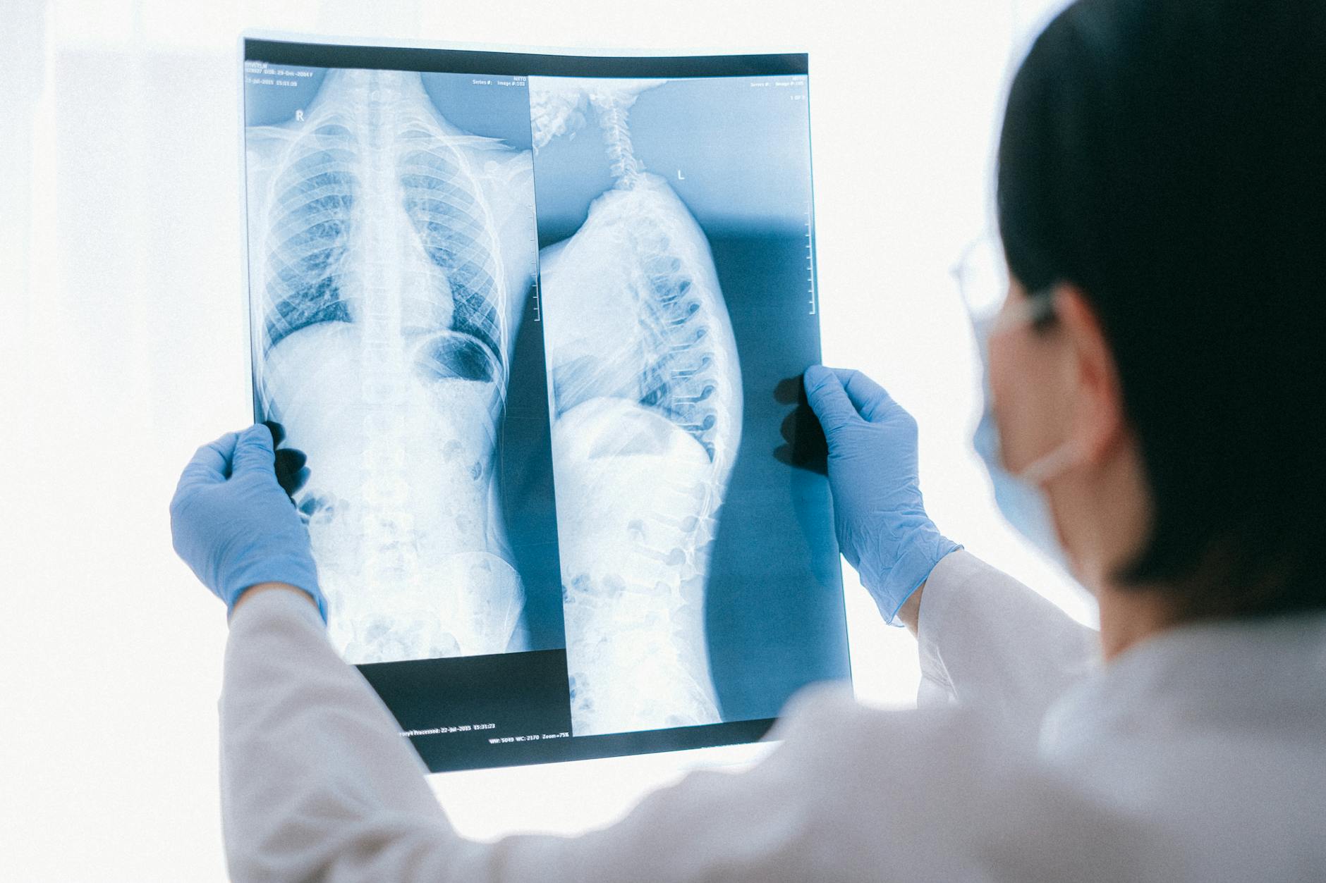
Medical research has documented that early-stage dementia frequently presents with subtle or undetectable changes on standard MRI, yet the symptoms and clinical evaluation clearly point to dementia. A patient in their 60s or early 70s might have a two-year history of word-finding difficulties, getting lost in familiar places, and changes in personality, yet their MRI shows no significant atrophy or pathology. The brain hasn’t visibly shrunk yet, the ventricles aren’t enlarged, and white matter disease appears minimal. However, neuropsychological testing reveals memory scores in the impaired range, and biomarkers suggest significant amyloid pathology is already present.
The critical limitation of relying solely on MRI in early dementia is that you can miss diagnoses altogether. A patient or family might come away thinking “the doctor said my scans are normal, so it must not be dementia.” In reality, the normal MRI says nothing about functional decline or underlying pathology. This can delay proper diagnosis by months or years, preventing early interventions that might slow progression. The normal-appearing brain can still be accumulating the toxic proteins that cause dementia, building toward the day when structural changes finally become visible.
How Dementia Diagnosis Really Works
A definitive dementia diagnosis cannot be made by MRI alone and, in fact, should never rely on imaging as the sole diagnostic tool. Instead, diagnosis requires a combination of brain scans, clinical symptoms observed by a neurologist or geriatrician, and cognitive testing administered by a neuropsychologist. The MRI provides supporting information—ruling out stroke, tumor, or other structural conditions that might cause cognitive symptoms—but it’s one piece among many.
A complete diagnostic workup includes standardized cognitive tests like the Montreal Cognitive Assessment or Mini-Cog, blood tests for biomarkers, review of the patient’s functional decline over time, and assessment of how symptoms affect daily life. A 58-year-old with a three-year history of forgetting appointments, inability to balance checkbooks, and mood changes might have normal MRI findings, but combined with poor performance on cognitive tests and family observations of decline, the clinical picture becomes clear: this is dementia, despite the normal scan. The normal MRI doesn’t negate the diagnosis; it just means the structural changes haven’t progressed far enough to be visible yet.

Biomarkers and Advanced Imaging When MRI Falls Short
When structural imaging is normal, biomarkers—chemical markers of dementia pathology in blood and cerebrospinal fluid—significantly improve diagnostic accuracy. Blood tests can now measure phosphorylated tau, amyloid beta ratios, and other markers that reflect brain pathology even when the brain still looks normal on MRI. These biomarkers are superior to diagnoses based solely on clinical evaluation and imaging, providing objective evidence of the underlying disease process.
PET (positron emission tomography) imaging is often more sensitive than MRI for dementia detection, particularly when MRI appears normal. FDG-PET scans can identify regions of reduced brain activity and detect amyloid beta plaque deposits, revealing functional problems that standard MRI misses entirely. While PET is more expensive and less routinely available than MRI, it can be invaluable when someone has clear dementia symptoms but normal structural imaging. The tradeoff is between cost and access versus diagnostic certainty—PET provides definitive evidence of amyloid pathology, but not every patient or healthcare system has easy access to this technology.
What MRI Cannot Diagnose
It’s essential to understand the fundamental limitation: MRI cannot directly diagnose dementia on its own, even if it’s abnormal. A brain scan might show some atrophy or white matter changes, but many people with these findings never develop dementia, while others with minimal visible changes develop severe cognitive decline. MRI is a supportive tool, useful for excluding other conditions (stroke, tumor, hydrocephalus), but it’s not diagnostic for dementia itself. The danger of over-relying on MRI is that patients and families might misinterpret results.
A radiologist’s report saying “mild age-appropriate atrophy” might seem reassuring but doesn’t rule out dementia. Conversely, someone with obvious dementia symptoms might receive a normal MRI report and feel falsely reassured that nothing is wrong. This is why a multi-pronged diagnostic approach—combining imaging, cognitive testing, biomarkers, and clinical assessment—is the standard of care. No single test should be used to confirm or exclude dementia.
Advanced MRI Techniques Improving Early Detection
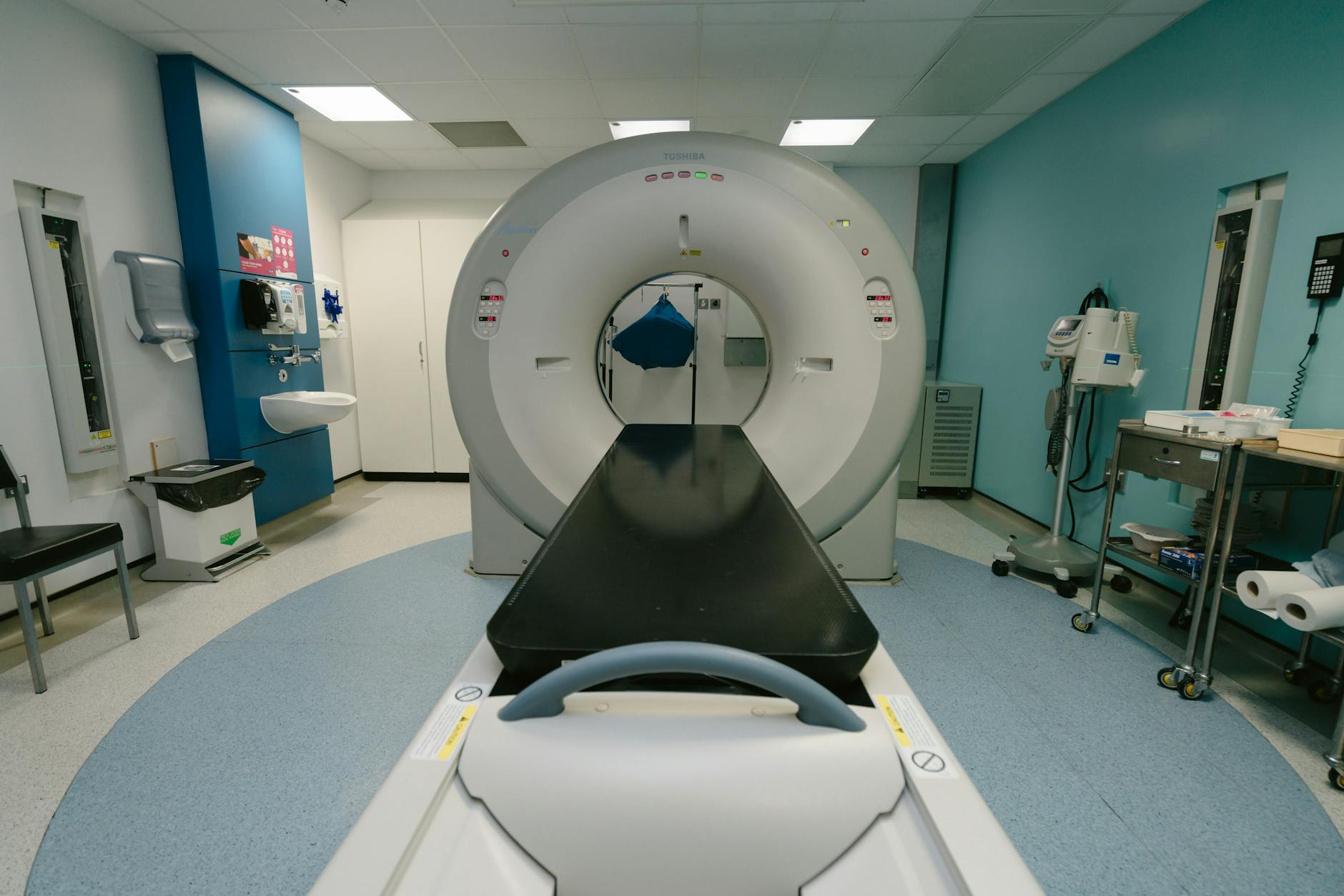
Recent advances in brain imaging technology are improving our ability to detect dementia earlier, even before standard MRI shows obvious changes. High-field MRI machines operating at 3 Tesla (3T) with specialized sequences can identify subtle hippocampal atrophy and white matter changes that conventional MRI might miss. Semi-quantitative visual scales and volumetric analysis of specific brain regions provide more sensitive measurement of the subtle shrinkage that occurs in early dementia.
These advanced techniques are still primarily used in research settings and specialized memory clinics, but they represent the direction of future diagnostic capability. A 72-year-old with early cognitive complaints might have a normal conventional MRI, but a 3T machine with specialized hippocampal protocols could detect the millimeter-level shrinkage characteristic of early Alzheimer’s disease. As these techniques become more widely available, diagnosis will shift toward earlier detection, potentially before symptoms become severe enough to significantly impact daily life.
What This Means for Patients and Families Looking Ahead
The future of dementia diagnosis is moving away from reliance on any single imaging test and toward integrated assessment combining multiple sources of information. Blood biomarkers are becoming more refined and accessible, PET imaging is being standardized, and advanced MRI protocols are transitioning from research to clinical practice. This multipronged approach will catch dementia earlier and more reliably, even in patients who might have had normal conventional MRI findings years ago.
For patients and families right now, this means understanding that a normal MRI doesn’t provide reassurance or exclude dementia. It’s a piece of information that needs to be interpreted within the full clinical context. If someone is experiencing cognitive decline, pursuing comprehensive neuropsychological testing, biomarker evaluation, and specialist assessment is essential, regardless of what the MRI shows.
Conclusion
The reality that dementia can exist with a normal brain MRI reflects an important truth about how this disease develops: pathology and symptoms often precede visible structural change. Early dementia is fundamentally a functional and molecular disease before it becomes a structural one. A normal MRI doesn’t mean the brain is healthy or that cognitive decline isn’t real; it means the visible damage hasn’t accumulated enough to show up on standard imaging. This doesn’t make the dementia less serious or the cognitive changes less real—it makes early diagnosis more challenging but also more crucial, because interventions are most effective in the earliest stages.
If you or a loved one is experiencing cognitive changes, insist on comprehensive evaluation that goes beyond MRI. Request cognitive testing, blood biomarker assessment, and specialist evaluation by a neurologist or memory specialist. A normal brain scan should prompt more thorough investigation, not reassurance that nothing is wrong. The dementia diagnosis of today requires a complete clinical picture, combining imaging, testing, biomarkers, and expert assessment—and families deserve care that reflects this modern understanding.