People diagnosed with Alzheimer’s disease lose roughly 1.9% of their total brain volume each year, nearly four times the rate seen in healthy aging. To put that in perspective, a healthy 65-year-old loses about 0.5% of brain volume annually, while someone with Alzheimer’s at the same age may be losing tissue at a pace that compounds into devastating cognitive decline within just a few years. The hippocampus, the brain’s memory hub, takes the hardest hit, shrinking at an average rate of 4.66% per year in Alzheimer’s patients compared to about 1.41% per year in normal aging.
These numbers come from decades of MRI research, and they matter far beyond the academic realm. Structural brain imaging has become one of the most reliable ways to track disease progression, differentiate Alzheimer’s from normal aging, and even detect changes years before a clinical diagnosis. This article walks through what MRI studies reveal about annual brain volume loss at each stage of cognitive decline, which regions deteriorate first, how new imaging technology is changing detection, and what recent drug trials have unexpectedly shown about the relationship between treatment and brain shrinkage.
Table of Contents
- How Much Brain Volume Do Alzheimer’s Patients Lose Per Year Compared to Normal Aging?
- Why the Hippocampus Loses Volume Faster Than Any Other Brain Region
- The Predictable Spread of Atrophy Across Brain Regions
- How Quantitative MRI Technology Is Changing Early Detection
- The Paradox of Brain Shrinkage in Anti-Amyloid Drug Trials
- What Six Months of MRI Data Can Reveal About Prognosis
- Where Brain Volumetric Research Is Heading
- Conclusion
- Frequently Asked Questions
How Much Brain Volume Do Alzheimer’s Patients Lose Per Year Compared to Normal Aging?
The difference between normal age-related brain shrinkage and Alzheimer’s-driven atrophy is stark once you look at the numbers side by side. Healthy adults begin losing brain volume gradually after age 35, at a rate of about 0.2% per year. By age 60, that rate accelerates to roughly 0.5% per year, which translates to about 2 to 2.4% per decade. This is the biological baseline, and most people never notice it in their daily functioning. In Alzheimer’s disease, however, whole-brain atrophy averages 1.9% per year, with moderate-stage patients sometimes reaching 2 to 3% annually. That three- to four-fold acceleration is not subtle, and it maps directly to the cognitive losses that families witness at the bedside. Mild cognitive impairment, often considered a transitional stage between normal aging and dementia, falls right in the middle.
MCI patients lose approximately 1.2% of whole-brain volume per year. Not everyone with MCI progresses to Alzheimer’s, but those who do tend to show atrophy rates that creep closer to the Alzheimer’s range over time. This is one reason serial MRI scans, taken six to twelve months apart, have become a valuable clinical tool. A single scan can show that a brain looks smaller than expected for age, but two scans separated by time reveal whether the rate of loss is following the normal track or something far more aggressive. Consider a 72-year-old woman whose first MRI shows mild hippocampal atrophy. On its own, that finding could reflect normal aging. But a follow-up scan twelve months later showing nearly 5% hippocampal volume loss puts her squarely in the range associated with Alzheimer’s disease, not healthy aging. This kind of longitudinal comparison is where MRI data shifts from interesting to clinically actionable.
Why the Hippocampus Loses Volume Faster Than Any Other Brain Region
The hippocampus is ground zero for Alzheimer’s-related atrophy, and the numbers reflect just how disproportionately it suffers. A meta-analysis pooling data from multiple MRI studies found that Alzheimer’s patients lose an average of 4.66% of hippocampal volume per year, with a 95% confidence interval of 3.92 to 5.40%. mci patients lose approximately 2.5% per year, while healthy controls lose about 1.41%. By the time Alzheimer’s is clinically diagnosed, the hippocampus has typically already shrunk by roughly 24.2% on the left side and 23.1% on the right compared to age-matched controls. In advanced stages, the rate of hippocampal shrinkage can reach 10 to 15% per year, a pace that leaves little functional tissue behind. This vulnerability makes sense biologically. The hippocampus is one of the first regions where amyloid plaques and tau tangles accumulate, and it is critical for forming new memories.
The earliest symptom most families notice, the repetition of questions or forgetting recent conversations, maps directly to hippocampal dysfunction. However, hippocampal atrophy alone does not confirm Alzheimer’s. Other conditions, including frontotemporal dementia, chronic depression, post-traumatic stress disorder, and even prolonged high-dose corticosteroid use, can cause hippocampal shrinkage. Clinicians must interpret volume loss in context, alongside biomarker data, cognitive testing, and clinical history. An isolated hippocampal measurement without that broader picture can lead to misattribution. It is also worth noting that researchers have detected significant hippocampal volume loss on MRI in as little as six months in patients with MCI and Alzheimer’s disease. This sensitivity makes the hippocampus a particularly useful target for monitoring, but it also means that even short delays in follow-up imaging can miss the window where intervention might still have meaningful impact.
The Predictable Spread of Atrophy Across Brain Regions
alzheimer‘s does not attack the brain randomly. MRI studies have mapped a remarkably consistent pattern of regional progression, and understanding this sequence has practical implications for diagnosis and staging. Atrophy begins in the medial temporal lobes and the fusiform gyrus, a region involved in facial recognition and visual processing, at least three years before a clinical diagnosis is made. This means the brain is already losing measurable tissue volume while the person may still be passing standard cognitive screening tests. From the medial temporal lobes, atrophy spreads to the posterior temporal and parietal lobes, which govern language comprehension, spatial orientation, and the ability to integrate sensory information.
This is typically when symptoms become obvious enough for families to seek medical attention. Problems like getting lost in familiar neighborhoods, difficulty managing finances, or struggling to follow conversations correspond to tissue loss in these regions. In later stages, the frontal lobes are affected, bringing changes in personality, judgment, and executive function that often transform the caregiving experience entirely. For example, a patient who initially presents with pure memory complaints but whose MRI shows atrophy extending into the parietal lobes is likely further along in disease progression than cognitive test scores alone might suggest. This spatial information from imaging can help clinicians set more realistic expectations with families and plan care accordingly, rather than relying solely on bedside cognitive assessments that may underestimate true disease burden.

How Quantitative MRI Technology Is Changing Early Detection
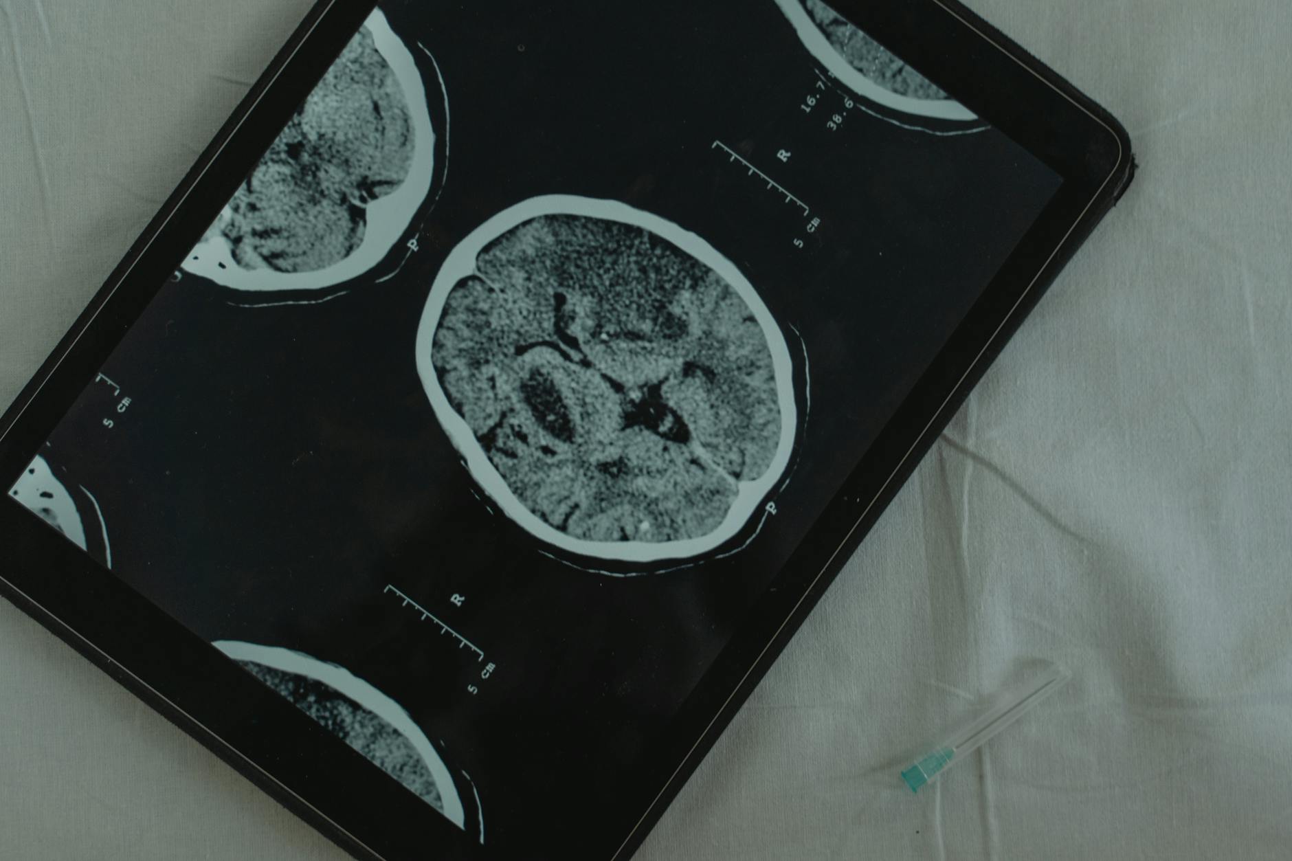
Traditional MRI reads are subjective. A radiologist looks at brain structures and judges whether they appear smaller than expected for age. This visual assessment catches moderate to severe atrophy reliably but often misses early-stage changes that fall within the range of what might look normal. Newer quantitative approaches are closing that gap. Tools like 3T NeuroQuant MRI now provide automated volumetric analysis, measuring the precise volume of dozens of brain structures and comparing them against age- and sex-matched norms. This turns a subjective visual impression into an objective number that can be tracked over time. An even larger step forward involves normative brain charts developed from data on 56,173 participants.
Much like pediatric growth charts that plot a child’s height and weight against population centiles, these brain charts allow clinicians to see where an individual patient falls relative to thousands of others of the same age and sex. A 68-year-old man whose hippocampal volume sits at the 8th percentile for his demographic is in a very different clinical situation than one at the 45th percentile, even if both are reporting mild forgetfulness. The tradeoff with quantitative imaging is accessibility and cost. Not every imaging center has 3T MRI capability or NeuroQuant software, and insurance coverage for volumetric analysis remains inconsistent. Many community hospitals still rely on 1.5T scanners and qualitative reads. Patients in rural areas or those without strong advocacy from their physicians may not receive the kind of imaging that could detect early changes. The technology exists, but its distribution lags well behind its capability.
The Paradox of Brain Shrinkage in Anti-Amyloid Drug Trials
One of the most counterintuitive findings in recent Alzheimer’s research involves the newest class of treatments. Anti-amyloid antibodies like donanemab and lecanemab, which clear amyloid plaques from the brain, showed a clinical benefit in slowing cognitive decline. But the MRI data told a puzzling story. Patients receiving these drugs actually lost more brain volume than those on placebo at both 52 and 76 weeks of treatment. This was the opposite of what researchers expected. If the drugs were working, shouldn’t the brain be shrinking less? Several theories attempt to explain this paradox. One is that removing large deposits of amyloid physically reduces the volume of tissue, since plaques themselves take up space.
Another is that the inflammatory response triggered by amyloid clearance causes transient fluid shifts or tissue changes that register as volume loss on MRI. Supporting this second theory is the fact that more than one in three patients treated with these drugs developed ARIA, amyloid-related imaging abnormalities, which include brain swelling and microbleeding visible on MRI. This finding creates a genuine clinical dilemma. Brain volume loss on MRI has long been used as a marker of disease progression. If an effective treatment actually accelerates volume loss in the short term, clinicians can no longer rely on serial MRI alone to judge whether a patient is responding to therapy. The clinical significance is still under investigation, and it serves as an important warning against interpreting any single biomarker in isolation. Families tracking a loved one’s treatment should be aware that more brain shrinkage on a follow-up scan does not necessarily mean the drug is failing.

What Six Months of MRI Data Can Reveal About Prognosis
One of the more practical findings from longitudinal MRI research is that meaningful changes in brain volume can be detected in as little as six months. This has shifted the clinical conversation about how often imaging should be repeated. In research settings, six-month intervals are common for tracking MCI patients who may be converting to Alzheimer’s.
In clinical practice, annual scans are more typical, though the optimal frequency remains a matter of professional judgment. For a patient diagnosed with MCI, a six-month follow-up MRI showing hippocampal loss in the 2.5% annual range provides stronger prognostic information than cognitive testing alone. It can help determine whether to initiate treatment earlier, connect the patient with clinical trials, or begin long-term care planning while the patient can still participate in those decisions. The scan does not change the disease itself, but it changes the timeline of the response.
Where Brain Volumetric Research Is Heading
The convergence of large-scale normative datasets, automated volumetric software, and machine learning models is pushing MRI-based detection toward true population-level screening. Normative brain charts built from tens of thousands of participants are being refined to account for genetic background, education level, and cardiovascular risk factors, all of which influence brain volume independently of Alzheimer’s pathology. The goal is a future where a routine brain MRI at age 55 or 60 generates an individualized risk profile, flagging people whose atrophy trajectory looks abnormal years before symptoms emerge. Whether that future arrives depends on factors beyond technology.
Reimbursement models, clinical workflow integration, and the question of what to do with early detection in the absence of a disease-modifying cure all remain unresolved. But the imaging science itself is no longer the bottleneck. The data showing that Alzheimer’s-related atrophy is detectable, quantifiable, and distinct from normal aging is robust. The challenge now is ensuring that knowledge reaches patients early enough to matter.
Conclusion
MRI research has established clear, reproducible benchmarks for brain volume loss across the spectrum of aging and Alzheimer’s disease. Healthy aging costs the brain about 0.5% of its volume per year after age 60. Mild cognitive impairment roughly doubles that rate to 1.2% per year. Alzheimer’s disease nearly quadruples it to 1.9% per year for whole-brain volume and drives hippocampal loss to 4.66% annually. These are not abstract numbers.
They correspond directly to the memory failures, disorientation, and personality changes that define the disease for the people living through it. For families and clinicians navigating a diagnosis, understanding these rates provides both a framework for what to expect and a basis for evaluating treatment options. Quantitative MRI is making early detection more precise, though access remains uneven. The unexpected findings from anti-amyloid drug trials remind us that brain volume is a powerful but imperfect proxy for disease activity. The most useful approach combines serial imaging with cognitive testing, biomarker analysis, and honest clinical judgment, none of which is sufficient on its own.
Frequently Asked Questions
How fast does the brain shrink in Alzheimer’s compared to normal aging?
Alzheimer’s patients lose about 1.9% of whole-brain volume per year, compared to approximately 0.5% per year in healthy older adults. The hippocampus, critical for memory, shrinks at 4.66% per year in Alzheimer’s versus about 1.41% per year in normal aging.
Can MRI detect Alzheimer’s before symptoms appear?
Yes. Research shows that atrophy in the medial temporal lobes and fusiform gyrus is detectable on MRI at least three years before a clinical diagnosis. Quantitative volumetric MRI tools like NeuroQuant can identify abnormal shrinkage patterns that visual reads might miss.
How often should someone with MCI get a brain MRI?
Research has shown that significant hippocampal volume loss can be detected in as little as six months. In clinical practice, annual scans are common for MCI patients, though your neurologist may recommend six-month intervals depending on the clinical picture.
Does the hippocampus shrink on both sides equally in Alzheimer’s?
Not exactly. Studies show the left hippocampus tends to lose slightly more volume, averaging about 24.2% reduction compared to 23.1% on the right when measured against healthy controls. Both sides are significantly affected, but the asymmetry is consistent across studies.
Do the new Alzheimer’s drugs like lecanemab stop brain shrinkage?
Paradoxically, no. Clinical trials of donanemab and lecanemab showed that treated patients actually lost more brain volume than placebo groups at 52 and 76 weeks. This may be related to amyloid removal or the high rate of brain swelling and bleeding seen in over a third of treated patients. The clinical meaning of this finding is still being studied.
What is a NeuroQuant MRI?
NeuroQuant is an FDA-cleared software tool used with 3T MRI scanners that automatically measures the volume of specific brain structures and compares them against normative databases adjusted for age and sex. It provides objective, numerical assessments rather than relying on a radiologist’s visual impression alone.