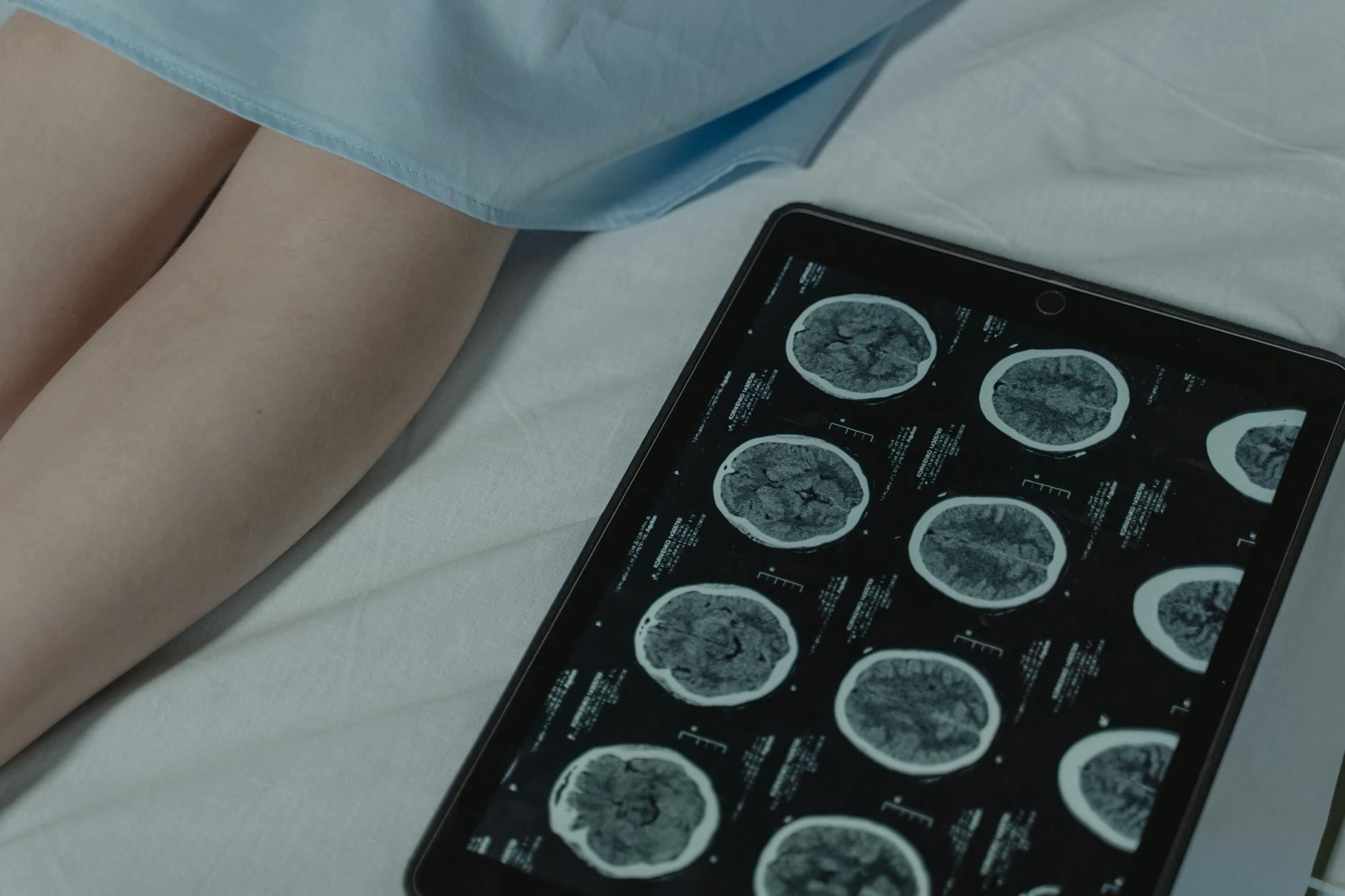
Brain imaging sits at the center of this dementia and brain health question.
Recent advances in brain imaging technology are fundamentally transforming Alzheimer’s disease screening, enabling clinicians to detect the disease at earlier stages—even before symptoms appear. A 2025 meta-analysis found that deep learning tools analyzing PET scans achieve 98% diagnostic accuracy, while blood-based biomarker tests like p-tau217 can now identify Alzheimer’s pathology across all disease stages, from pre-symptomatic to advanced dementia. The Alzheimer’s Association has formally recognized blood biomarker tests meeting specific accuracy thresholds (≥90% sensitivity and ≥90% specificity) as valid substitutes for invasive PET imaging or cerebrospinal fluid testing, fundamentally changing clinical practice.
For example, a patient experiencing mild memory concerns can now be evaluated through a simple blood test rather than requiring a positron emission tomography scan or lumbar puncture, reducing both cost and patient burden while delivering more reliable results. These imaging and biomarker advances represent a new era in Alzheimer’s screening. Rather than waiting for cognitive decline to become obvious, healthcare providers can now identify brain changes that precede symptoms by years or even decades. This article explores how these technological breakthroughs work, their accuracy compared to traditional methods, their practical applications in clinical settings, and the limitations patients and families should understand.
Table of Contents
- How Advanced Brain Imaging Detects Alzheimer’s Disease Earlier Than Symptoms Appear
- Blood-Based Biomarkers: The Non-Invasive Revolution in Alzheimer’s Detection
- Combining Imaging Modalities for Superior Diagnostic Accuracy
- AI and Deep Learning: Enhancing Interpretation of Brain Images
- Emerging Technologies: Optical Imaging and Novel Detection Methods
- Implementing Advanced Screening Programs in Clinical Practice
- The Path Forward: From Detection to Prevention and Treatment
- Conclusion
How Advanced Brain Imaging Detects Alzheimer’s Disease Earlier Than Symptoms Appear
alzheimer‘s disease begins long before memory loss becomes noticeable. The hallmark pathologies—amyloid-beta plaques and tau tangles—accumulate silently in the brain for 10 to 20 years before cognitive symptoms emerge. Modern brain imaging captures these pathological changes, giving clinicians a window into disease progression before damage becomes irreversible. PET imaging, specifically 18F-FDG PET/CT, demonstrates this power quantitatively. Studies show that 18F-FDG PET/CT achieves 92% sensitivity and 78% specificity when distinguishing Alzheimer’s disease from other dementias. Amyloid PET scans are even more selective, with greater than 90% sensitivity and greater than 70% specificity for Alzheimer’s diagnosis.
Tauvid, an FDA-approved tau PET tracer introduced in 2020, enabled clinicians to visualize tau pathology directly in living patients for the first time, providing unprecedented specificity for Alzheimer’s-related changes. However, PET imaging requires access to specialized centers, exposes patients to radioactive tracers, and often costs thousands of dollars per scan—making it impractical for universal screening programs. MRI offers a more accessible alternative, detecting structural brain changes without radiation. Hippocampal volume (the seahorse-shaped region crucial for memory) shrinks as Alzheimer’s progresses. Across 33 studies involving approximately 4,000 subjects, MRI-based hippocampal measurement achieved 73% pooled sensitivity and 71% specificity for distinguishing Alzheimer’s from controls. While these performance metrics are respectable, they fall short of PET imaging’s precision, meaning some early-stage patients may be missed using MRI alone.

Blood-Based Biomarkers: The Non-Invasive Revolution in Alzheimer’s Detection
The most significant recent breakthrough in Alzheimer’s screening is the validation of blood-based biomarkers that detect Alzheimer’s pathology from a simple blood draw. Unlike PET scans or CSF testing, blood tests require no special equipment, radiation exposure, or invasive procedures, making them genuinely feasible for population-wide screening. P-tau217 has emerged as the most promising biomarker, with studies confirming its ability to accurately detect Alzheimer’s pathology at all disease stages—pre-symptomatic, mild cognitive impairment, and dementia. According to the 2025 NIH Alzheimer’s Disease and Related Dementias Research Progress Report, p-tau217 blood tests now rival imaging modalities in precision while remaining non-invasive and cost-effective.
The test can be performed in routine clinical laboratories, primary care offices, or screening programs, removing the geographic and logistical barriers that have historically limited Alzheimer’s diagnosis to specialized memory clinics. Two important limitations warrant mention: first, while blood biomarkers identify Alzheimer’s pathology, they do not diagnose cognitive symptoms—some individuals with Alzheimer’s brain pathology never develop dementia in their lifetime. Second, if a patient has cognitive symptoms but negative biomarkers, additional investigation may be necessary to identify non-Alzheimer’s causes. Blood biomarkers are most valuable when combined with cognitive assessment and used in structured screening protocols rather than in isolation. Additional validated blood biomarkers include p-tau181, p-tau231, and tau and amyloid pathophysiology markers, giving clinicians a portfolio of tests to choose from based on clinical context and available resources.
Combining Imaging Modalities for Superior Diagnostic Accuracy
No single imaging technique captures the entire picture of Alzheimer’s disease. While individual modalities—MRI, PET, or blood biomarkers—perform well in isolation, combining multiple approaches yields significantly improved diagnostic accuracy and confidence in results. Recent research demonstrates that combining MRI and PET imaging substantially outperforms either modality alone for early Alzheimer’s detection. advanced ensemble learning methods that integrate information from both structural MRI (showing brain atrophy patterns) and functional PET data (showing metabolic abnormalities) achieve diagnostic accuracy exceeding 95% in research settings. A typical multimodal screening might begin with MRI to assess hippocampal volume and regional atrophy, followed by blood biomarker testing to detect pathological proteins, and if results are discordant or inconclusive, confirmatory PET imaging.
This stepwise approach optimizes both cost-effectiveness and diagnostic precision. For instance, an older adult with subjective cognitive decline might start with a blood biomarker test (inexpensive, non-invasive) combined with MRI (more accessible than PET); if findings suggest Alzheimer’s pathology, amyloid or tau PET imaging can confirm the diagnosis before initiating preventive treatments. The multimodal approach also accounts for individual variation in how Alzheimer’s manifests. Some patients show prominent amyloid accumulation with minimal tau pathology, while others demonstrate the reverse. Others have “atypical” presentations with asymmetric patterns of brain atrophy that don’t fit the classic description. Combining multiple imaging techniques reveals these individual patterns, guiding personalized treatment decisions.
AI and Deep Learning: Enhancing Interpretation of Brain Images
Artificial intelligence is dramatically improving how clinicians interpret brain imaging data. Deep learning algorithms trained on thousands of labeled brain scans can identify subtle pathological changes that human radiologists might overlook, and they process images consistently without fatigue or interpretive drift. A 2025 meta-analysis of deep learning tools for Alzheimer’s diagnosis using PET scans reported a pooled Area Under the Curve (AUC) of 98%—a level of diagnostic performance that approaches perfection. These AI systems excel at detecting patterns across high-dimensional imaging data, identifying combinations of regional changes that indicate Alzheimer’s pathology.
In practical clinical settings, AI interpretation systems assist radiologists by flagging suspicious regions, prioritizing abnormal scans, and providing second-opinion analysis. A radiologist reviewing 50 MRI scans from a memory clinic might miss subtle hippocampal atrophy in one or two cases due to fatigue; an AI system analyzing the same scans would identify pathology consistently. However, AI interpretation systems require extensive validation in the specific population and imaging protocols used at each institution, and they perform best when radiologists remain engaged in the interpretive process rather than accepting AI predictions blindly. The integration of AI into clinical workflow also enables faster turnaround on scan interpretation, reducing the weeks patients typically wait for results. In research environments, AI-enhanced image analysis has enabled large population-based studies of Alzheimer’s preclinical pathology that would have been logistically impossible using human interpretation alone.
Emerging Technologies: Optical Imaging and Novel Detection Methods
Beyond conventional PET and MRI, researchers are developing entirely new approaches to detect Alzheimer’s-related brain changes. One particularly promising innovation is optical spectroscopy—a non-invasive technique that uses light to identify structural changes in brain tissue without radiation or special equipment. Researchers at the Department of Veterans Affairs Bedford and Boston health systems recently developed an optical spectroscopy technique capable of identifying Alzheimer’s-related brain structural changes non-invasively through the scalp. This emerging technology holds potential for simple, inexpensive point-of-care screening in primary care settings or community health centers.
However, optical imaging remains largely experimental; its clinical performance has not yet been validated across large patient populations, and its advantages over blood biomarkers for practical screening applications remain uncertain. The technology also requires specialized optical equipment that is not yet commercially available or integrated into standard clinical practice. These novel approaches highlight the rapid pace of innovation in Alzheimer’s diagnostics. Within the next 5 to 10 years, clinicians may have access to screening tools that are simultaneously non-invasive, inexpensive, portable, and highly accurate—capabilities that could enable true population-wide early detection programs rather than selective screening in specialty clinics.

Implementing Advanced Screening Programs in Clinical Practice
For clinicians and healthcare systems considering how to integrate advanced imaging and biomarkers into their practice, the key challenge is establishing a rational, cost-effective screening pathway rather than ordering all available tests indiscriminately. The Alzheimer’s Association’s updated clinical practice guidelines provide guidance: blood biomarker tests with ≥90% sensitivity and ≥90% specificity can now substitute for PET imaging or cerebrospinal fluid testing in appropriate clinical contexts. A practical screening algorithm might follow this sequence: First, cognitive assessment and clinical history identify candidates for biomarker testing (those with cognitive complaints or at genetic risk).
Second, blood biomarker testing (p-tau217, p-tau181, or similar) screens for Alzheimer’s pathology. Third, if biomarkers are positive and additional confirmation is needed, structural MRI assesses the degree of brain atrophy. Finally, if clinical diagnosis remains uncertain despite positive biomarkers, amyloid or tau PET imaging can provide definitive confirmation. This stepwise approach reduces unnecessary testing, controls costs, and reserves expensive PET imaging for cases where it adds diagnostic value—typically when results of earlier tests are contradictory or when clinical presentation is atypical.
The Path Forward: From Detection to Prevention and Treatment
The significance of these imaging advances extends far beyond diagnosis. Earlier detection of Alzheimer’s pathology creates opportunities for prevention and disease modification that were unavailable when symptoms were the only clinical indicator. Several anti-amyloid monoclonal antibodies have demonstrated modest slowing of cognitive decline when administered to patients with mild cognitive impairment or mild dementia who have confirmed amyloid pathology.
These drugs require biomarker confirmation of amyloid presence before initiation, making accurate, accessible biomarker testing essential for treatment access. As more disease-modifying therapies enter the pipeline—including anti-tau agents and therapies targeting neuroinflammation—the role of early detection becomes even more critical. Patients identified before symptom onset represent a unique population in which preventive therapies might be even more effective than treatments started after cognitive decline. The convergence of improved detection with emerging treatments creates a compelling case for systematic Alzheimer’s screening programs, particularly among at-risk populations including individuals over age 65, those with cognitive complaints, and carriers of the APOE4 genetic risk factor.
Conclusion
Brain imaging and biomarker advances have fundamentally improved Alzheimer’s detection, enabling clinicians to identify disease pathology earlier and with greater accuracy than ever before. Blood-based biomarkers now rival imaging modalities in precision while being more accessible and less invasive; deep learning tools interpret complex imaging data with near-perfect accuracy; and emerging technologies promise even more transformative capabilities in the coming years.
For patients and families, these advances mean that subjective memory concerns can now be evaluated definitively through non-invasive testing, and early detection creates opportunities for disease-modifying treatments not available to those diagnosed after symptoms appear. If you or a family member are experiencing cognitive concerns, discussing biomarker testing with your primary care provider or a memory care specialist represents a logical first step. Early evaluation using these advanced techniques can provide clarity about whether cognitive changes reflect normal aging, Alzheimer’s pathology, or other treatable conditions—information that becomes increasingly valuable as evidence-based preventive and treatment options expand.
You Might Also Like
- Scientists Explain How Existing Alzheimer’s Drug Clears Brain Plaques
- Brain Volume Data Supports Efficacy of Oral Alzheimer’s Drug
- Brain Cell Communication Pathway Identified as Alzheimer’s Defense Mechanism
For more, see Alzheimer’s Association — caregiving.