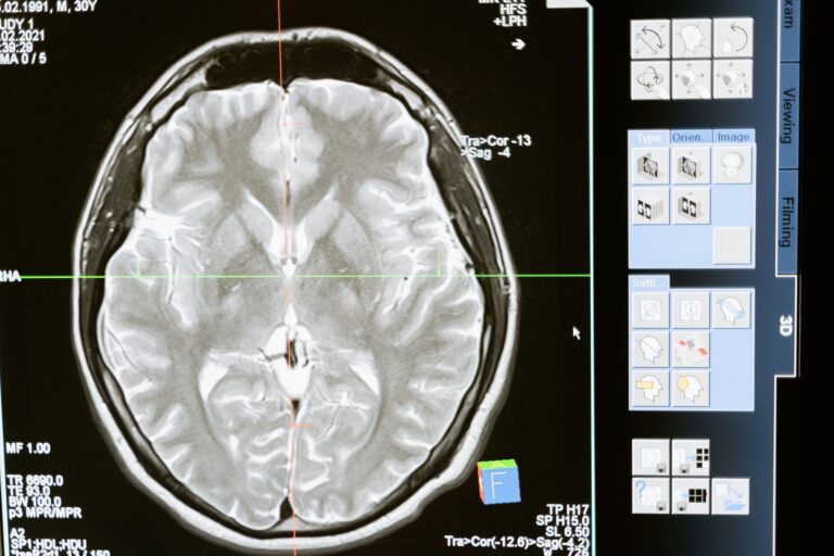
A structural brain MRI before an Alzheimer's evaluation can support diagnosis and rule out other causes, but it cannot diagnose Alzheimer's by itself. Structural MRI is a detailed magnetic scan that shows shrinkage patterns and blood-vessel damage in the brain. Shrinkage may support Alzheimer's or another neurodegenerative dementia, but it cannot specify which diagnosis applies. The scan also helps doctors track memory and thinking changes over time with repeat scans.
Medical information disclaimer: This article is for general educational purposes only and does not provide medical advice, diagnosis, or treatment. Always consult a physician or other qualified health professional about symptoms, medications, tests, or treatment decisions.
Table of Contents
- What a brain MRI can reveal
- What MRI cannot prove
- When treatment makes MRI required
- Safety, comfort, and trade-offs
What a brain MRI can reveal
Structural MRI shows atrophy patterns and detailed small-vessel disease. According to the National Institute on Aging, this shrinkage can support Alzheimer's or another dementia but cannot identify the exact type on its own NIA biomarker guidance. A standard workup often includes MRI or CT to support diagnosis and exclude other causes.
According to the National Institute on Aging, repeat scans also help track changes in memory and cognition Alzheimer's diagnostic guidelines. MRI also helps rule out treatable conditions that need different care. These mimics include brain tumor, stroke, bleeding, and normal-pressure hydrocephalus, alongside atrophy or small-stroke damage.
What MRI cannot prove
Conventional MRI is nonspecific for Alzheimer's because it does not directly show amyloid plaques or tau tangles. The 2024 Alzheimer's Association criteria define the disease biologically through Core 1 markers such as amyloid PET, approved spinal-fluid tests, and accurate plasma p-tau217. Early diagnostic accuracy is limited, especially in mild cognitive impairment.
Mild cognitive impairment means noticeable memory or thinking problems that do not yet prevent daily activities. One systematic review reported hippocampal MRI sensitivity around 0.62 and specificity 0.70. The reviewers found insufficient evidence for routine MRI to diagnose early dementia.
When treatment makes MRI required
Patients starting lecanemab, the FDA-approved anti-amyloid drug sold as Leqembi, must have a recent baseline brain MRI. Follow-up MRIs are scheduled during treatment to detect amyloid-related imaging abnormalities with swelling or bleeding. The schedule now includes a scan before the third infusion plus later infusions.
According to the FDA safety communication, these scans are meant to catch edema or hemorrhage early FDA drug safety communication. This monitoring role is different from diagnosis. Here MRI protects safety during therapy rather than confirming amyloid itself.
- Ask whether a recent baseline MRI is already on file
- Confirm the infusion-linked MRI schedule before starting
- Report headache, confusion, dizziness, vision changes, or weakness promptly
Safety, comfort, and trade-offs
MRI uses no ionizing radiation and gives better soft-tissue contrast than CT. That makes brain structures easier to distinguish without radiation exposure. FDA notes risks including projectile or device movement, thermal burns, implant heating, and claustrophobia.
Gadolinium contrast can be retained in the body and requires screening and benefit-risk assessment. Tell the imaging team about implants, pacemakers, aneurysm clips, cochlear devices, metal fragments, kidney problems, pregnancy, and prior contrast reactions. Ask about ear protection, how long you must lie still, whether contrast is needed, and what support is available for claustrophobia or back pain.