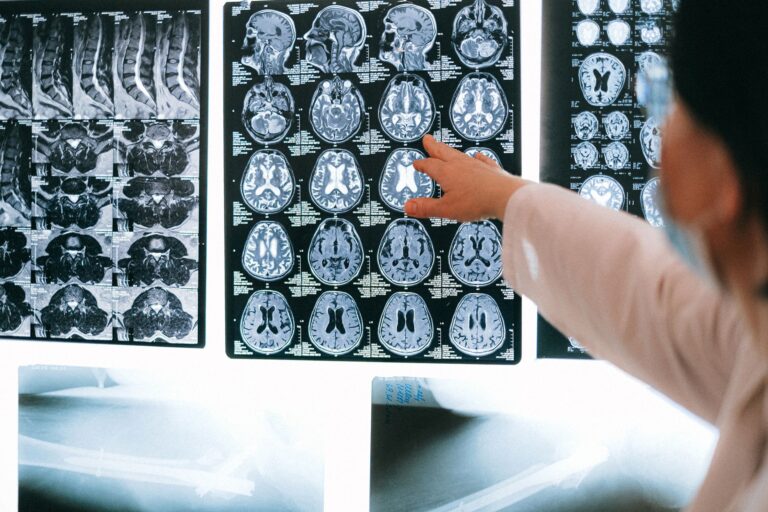
Amyloid PET and tau PET are two different brain scans that detect two different hallmarks of Alzheimer’s disease. An amyloid PET scan finds sticky beta-amyloid plaques that build up in the spaces between neurons, often years or even decades before symptoms appear. A tau PET scan detects abnormal tangles of tau protein that form inside neurons and tend to track much more closely with the actual location and severity of memory loss and thinking problems. Put simply: amyloid PET tells you whether the disease process has started, while tau PET tells you how far it has progressed and where it is causing damage. Consider two people, both with mild memory complaints.
One gets an amyloid PET scan that lights up positive, confirming that plaques are present, but the doctor still cannot say how quickly decline will come. A tau PET scan on that same person might show tau confined to a small region of the temporal lobe, suggesting early disease, or spread widely across the cortex, suggesting a more advanced and faster-moving process. The two scans answer related but distinct questions, which is why researchers increasingly use them together rather than treating one as a replacement for the other. Both are specialized nuclear medicine scans that use a small amount of radioactive tracer and a PET camera. Neither is a routine test you will get at an ordinary checkup, and in clinical practice amyloid PET is far more established and available than tau PET, which remains largely a research and specialty tool. Understanding what each one actually measures helps explain why a doctor might order one, both, or neither.
Medical information disclaimer: This article is for general educational purposes only and does not provide medical advice, diagnosis, or treatment. Always consult a physician or other qualified health professional about symptoms, medications, tests, or treatment decisions.
Table of Contents
- What Does Each Alzheimer’s Scan Detect, Amyloid PET or Tau PET?
- How Amyloid PET Plaque Imaging Works and Where It Falls Short
- How Tau PET Tangle Imaging Tracks Disease Progression
- Choosing Between Amyloid PET and Tau PET in Practice
- Limitations, Risks, and Misunderstandings of Both Scans
- Why Doctors Increasingly Use Amyloid and Tau Together
- What These Scans Mean for Newer Alzheimer’s Treatments
- Frequently Asked Questions
What Does Each Alzheimer’s Scan Detect, Amyloid PET or Tau PET?
The core difference comes down to which protein each scan is designed to find. Amyloid PET uses tracers such as florbetapir, florbetaben, or flutemetamol that bind to beta-amyloid plaques. These plaques accumulate outside neurons and are considered one of the earliest measurable changes in Alzheimer’s, sometimes present 15 to 20 years before any noticeable symptoms. A positive amyloid scan confirms plaque is present; a negative scan makes Alzheimer’s a very unlikely cause of someone’s memory problems. Tau PET, by contrast, uses tracers such as flortaucipir to detect neurofibrillary tangles of tau protein that form inside neurons.
Tau pathology matters because it correlates more tightly with cognitive symptoms than amyloid does. Amyloid can be widespread in the brain while a person still functions relatively well, but tau tends to appear in regions where neurons are actively dying, which is why its distribution often mirrors the specific problems a person is experiencing, whether that is memory, language, or spatial navigation. A useful comparison is to think of amyloid as smoke and tau as fire. Amyloid signals that something is wrong and that the Alzheimer’s process is underway, but the presence of smoke alone does not tell you how much is burning. Tau shows where the fire is actively consuming tissue. This is why a person can have a strongly positive amyloid scan yet only mild symptoms, while the extent of tau on a scan lines up more predictably with how impaired that person is.
How Amyloid PET Plaque Imaging Works and Where It Falls Short
An amyloid PET scan involves injecting the tracer into a vein, waiting a period for it to distribute and clear from the brain, and then lying still in the scanner while images are captured. A trained reader interprets the scan as either positive or negative based on whether the tracer signal appears in the cortex in a pattern consistent with plaque. In the United States, amyloid PET has clearer regulatory approval and more clinical experience behind it than tau PET, and it is often used to rule Alzheimer’s in or out when the diagnosis is uncertain. The major limitation of amyloid PET is that a positive result is common in older adults who do not have dementia. Amyloid plaques accumulate with normal aging, and a meaningful share of cognitively healthy people in their 70s and 80s will have a positive scan.
This means amyloid PET cannot, on its own, confirm that Alzheimer’s is the cause of a specific person’s symptoms. A positive scan in someone with memory complaints raises the probability of Alzheimer’s, but it does not prove causation, and over-reliance on it can lead to premature or incorrect diagnoses. There is also a practical warning worth noting. Amyloid PET has historically been expensive and often not covered by insurance for routine use, though coverage policies have been shifting. Ordering the scan without a clear clinical question can produce a result that confuses rather than clarifies, particularly when a patient interprets a positive scan as an immediate dementia diagnosis when it may reflect nothing more than age-related plaque with no functional consequence yet.
How Tau PET Tangle Imaging Tracks Disease Progression
Tau PET follows a similar procedure of tracer injection, uptake time, and scanning, but the tracer binds to tau tangles inside neurons instead of amyloid plaques. What makes tau PET distinctive is that the spread of tau through the brain tends to follow a fairly predictable staging pattern, beginning in the medial temporal lobe near the hippocampus and moving outward into wider cortical regions as disease advances. Because of this, a tau scan can give a rough sense of how far along the process has moved. This staging quality is where tau PET shines in a way amyloid PET cannot.
For example, a person with early symptoms whose tau is limited to the entorhinal cortex and hippocampus is likely at an earlier point than someone whose tau has spread into the parietal and frontal lobes. In research settings, this has helped predict the pace of decline, since heavier and more widespread tau burden generally signals faster progression and more severe impairment ahead. Tau PET has also helped explain unusual presentations of Alzheimer’s. In posterior cortical atrophy, a variant where people lose visual processing before memory, tau PET often shows heavy tracer uptake in the back of the brain corresponding to those visual symptoms, even when memory regions are relatively spared. That kind of localization is something amyloid PET, which tends to appear diffusely, is not well suited to reveal.
Choosing Between Amyloid PET and Tau PET in Practice
For most patients today, the practical reality is that amyloid PET is the scan a specialist is more likely to order, while tau PET remains concentrated in academic medical centers and clinical trials. Amyloid PET answers the more common clinical question of whether Alzheimer’s pathology is present at all, which is often the deciding factor in whether to pursue Alzheimer’s-specific treatments or look for another cause of cognitive change entirely. The tradeoff is one of certainty versus availability. Tau PET arguably provides information that maps more closely onto symptoms and prognosis, but it is harder to access, less standardized in how results are read, and not routinely reimbursed outside of research.
A memory clinic weighing which scan to use, if any, has to balance what question needs answering against what is actually obtainable and affordable for that patient. In many cases the answer is that neither scan is necessary, because history, cognitive testing, and simpler tests already point clearly to a diagnosis. It is also worth comparing PET scans to the newer blood-based biomarkers that are entering practice, which can measure amyloid and tau-related proteins from a blood draw. These are far cheaper and easier than a PET scan and are changing how testing is sequenced, though they are generally used to decide who should go on to confirmatory imaging rather than to replace PET entirely. A blood test might flag likely amyloid pathology, and a PET scan might then confirm it before a treatment decision is made.
Limitations, Risks, and Misunderstandings of Both Scans
Both scans involve exposure to a small dose of radiation from the tracer, which is generally considered low risk but is not zero, and this is a reason not to order these scans casually. Neither scan is a standalone diagnosis. A PET result is one piece of evidence that must be interpreted alongside a person’s history, examination, and cognitive testing. Treating a scan result as the final word is a common and serious misunderstanding. A specific warning applies to how results are communicated. A positive amyloid scan in an asymptomatic person can cause significant anxiety despite telling that person very little about whether or when they will develop dementia.
Because amyloid can be present for years without symptoms, and because not everyone with amyloid progresses to dementia on any predictable timeline, disclosing such results without careful counseling can do real harm. This is one reason amyloid imaging in people without symptoms is largely confined to research settings with built-in support. Tau PET carries its own limitations. Some tau tracers show off-target binding, meaning they can stick to structures unrelated to Alzheimer’s tau, which complicates interpretation. Tau tangles also occur in other conditions beyond Alzheimer’s, so a tau signal is not automatically specific to one disease. These technical and biological caveats are part of why tau PET has not yet moved into everyday clinical use the way amyloid imaging has.
Why Doctors Increasingly Use Amyloid and Tau Together
Rather than picking a winner, much of the research field now views amyloid and tau as complementary readouts of the same disease at different stages. The prevailing model holds that amyloid accumulates first and may help trigger or accelerate the spread of tau, while tau is the change that more directly drives neuron loss and symptoms. Seeing both gives a fuller picture than either alone.
In practice, this combined view underlies how some Alzheimer’s clinical trials select and monitor participants. A trial testing an anti-amyloid drug, for instance, might use amyloid PET to confirm eligibility and tau PET to gauge how advanced each participant’s disease is, since people with lower tau burden sometimes respond differently than those with extensive tangles already present. That pairing helps researchers interpret whether a treatment is actually changing the disease course.
What These Scans Mean for Newer Alzheimer’s Treatments
The arrival of anti-amyloid antibody treatments has made these scans more clinically relevant, because such drugs are generally intended for people with confirmed amyloid pathology. Confirming amyloid presence before starting these medications is important, since the treatments carry real risks, including brain swelling and small bleeds that require monitoring, and giving them to someone without amyloid would expose that person to risk with no expected benefit.
Tau imaging is increasingly discussed as a way to identify who is most likely to benefit and who may already be too advanced for a given therapy to help meaningfully. A person with minimal tau spread might be a stronger candidate for an early intervention than someone whose tau has already extended across much of the cortex. This is an active area of investigation rather than settled practice, and the specific role each scan will play in treatment decisions continues to be worked out in specialty centers.
Frequently Asked Questions
Is amyloid PET or tau PET more accurate for diagnosing Alzheimer’s?
Neither is a standalone diagnosis. Amyloid PET is better at confirming whether Alzheimer’s pathology is present at all, while tau PET tracks more closely with actual symptoms and disease stage. They answer different questions.
Can I get a tau PET scan at a regular hospital?
Usually not. Tau PET remains largely confined to academic medical centers and research studies, while amyloid PET is more widely available in specialty and memory clinics.
Does a positive amyloid scan mean I have dementia?
No. Amyloid plaques accumulate with normal aging, and many cognitively healthy older adults have positive scans. A positive result raises the probability of Alzheimer’s but does not confirm it as the cause of symptoms.
Are these scans dangerous?
Both use a small dose of radioactive tracer, generally considered low risk but not zero. This is one reason they are not ordered routinely without a clear clinical question.
How do blood tests fit in with PET scans?
Newer blood-based biomarkers can flag likely amyloid or tau pathology cheaply and are increasingly used to decide who should go on to confirmatory PET imaging, rather than fully replacing the scans.