When the sacroiliac joint becomes inflamed, it triggers pain that radiates from the lower back and buttocks, sometimes traveling down one or both legs—a condition affecting an estimated 15-30% of people with chronic low back pain. Imagine waking up stiff, spending the first hour of your day struggling to move comfortably, then feeling sharp pain when you bend to tie your shoes or walk up stairs. This is the daily reality for many experiencing SI joint inflammation, a condition that disrupts movement patterns and quality of life but is highly treatable with proper intervention.
This article explores what happens physically when the SI joint becomes inflamed, why it occurs, how to recognize the symptoms, and what evidence-based treatment options can restore function and reduce pain. The sacroiliac joint is a small but critical joint connecting your lowest rib to your pelvis. When the tissue around it becomes inflamed—whether from arthritis, pregnancy, trauma, or repetitive strain—it causes localized and referred pain patterns that can feel disabling but are not usually permanent. The good news: approximately 95% of patients respond well to non-surgical treatment, with significant improvement in 1-3 months.
Table of Contents
- How the SI Joint Becomes Inflamed and What Physical Changes Occur
- Recognizing the Symptoms and Understanding Pain Patterns
- Primary Causes and Risk Factors That Lead to Inflammation
- How Inflammation is Diagnosed and Confirmed
- Treatment Options and Expected Recovery Timelines
- Prevention Strategies and Long-Term Management
- When SI Joint Inflammation Signals Systemic Disease and Requires Specialized Care
- Conclusion
How the SI Joint Becomes Inflamed and What Physical Changes Occur
The sacroiliac joint sits at the base of your spine where your lower vertebrae connect to your pelvis. It contains cartilage, synovial fluid for lubrication, and supporting ligaments that allow minimal but essential movement during walking, bending, and lifting. When inflammation develops, the synovial membrane swells, reducing the fluid that allows smooth gliding, and the surrounding ligaments become irritated and tense. This inflammation happens gradually in some cases (like osteoarthritis developing over years) and suddenly in others (like a fall or acute injury). Several disease processes cause this inflammation. Ankylosing spondylitis, rheumatoid arthritis, and psoriatic arthritis specifically target the SI joint because it’s a natural site for inflammatory arthritis to develop.
In pregnancy, hormonal changes loosen the ligaments supporting the SI joint to prepare for childbirth, but this also destabilizes the joint and creates inflammation—a particular concern for women because SI joint dysfunction is more common in women overall. Trauma like a car accident or fall, combined with repetitive activities like running or heavy lifting, can strain the joint beyond its tolerance, triggering inflammation that persists long after the initial injury. The inflammatory cascade is not immediate. Often, a small movement pattern dysfunction—one leg slightly shorter than the other, weak hip muscles, or imbalanced running stride—creates uneven stress on the joint over weeks or months. The cartilage begins to wear, the synovial membrane becomes irritated, and inflammation builds until pain becomes noticeable. However, if inflammation occurs suddenly after a specific trauma, the pain onset can be dramatic and disabling within hours.

Recognizing the Symptoms and Understanding Pain Patterns
SI joint inflammation produces a specific constellation of symptoms that distinguish it from other back conditions. Lower back pain is the most common presentation, often concentrated on one side slightly above the buttock. The pain may be sharp and stabbing, or it may be a deep, aching throb. Many people also experience buttock pain that radiates down one leg—sometimes described as a pain pattern similar to sciatica, though it’s mechanical rather than nerve-root compression, which is an important distinction because it changes treatment approach. Morning stiffness lasting more than one hour is a hallmark symptom of SI joint inflammation, especially when inflammatory arthritis is the underlying cause. Patients report severe stiffness upon waking that gradually improves with movement and activity.
Numbness, tingling, or weakness in the affected leg can occur when inflammation swells enough to irritate the nearby nerve roots, though true neurological symptoms are less common than pain. Some people report instability—a sensation that the joint feels “loose” or “unstable”—and difficulty with movements like standing on one leg or climbing stairs. The pain is often worse with sitting, bending forward, or lying on the affected side, and better with walking or lying flat. A crucial limitation to note: not all buttock pain or lower back pain originates from the SI joint. Genuine sciatica (compression of the sciatic nerve), hip arthritis, or lumbar spine issues produce similar pain patterns, which is why proper diagnosis matters before beginning treatment. Someone might spend months treating what they assume is SI joint pain when the actual problem lies elsewhere, delaying effective treatment.
Primary Causes and Risk Factors That Lead to Inflammation
Inflammatory arthritis represents a major cause of SI joint inflammation. Ankylosing spondylitis, in particular, frequently affects the SI joint early in the disease course, making SI joint pain sometimes the first warning sign of systemic autoimmune disease. Rheumatoid arthritis and psoriatic arthritis can also target this joint. In these cases, the inflammation is driven by the body’s immune system attacking joint tissues, not by mechanical wear or injury. Pregnancy is a unique cause affecting women specifically.
During pregnancy, hormonal changes—particularly relaxin hormone—loosen the ligaments supporting the SI joint to allow pelvic opening for childbirth. This destabilization, combined with increased abdominal weight shifting the center of gravity forward, creates tremendous stress on the SI joint. Many pregnant women develop SI joint pain in the second and third trimesters, and for some, the pain persists months or years after delivery. Mechanical causes are equally important: leg length discrepancy (even a small difference), previous lumbar spine surgery, repetitive strain from running or heavy labor, and poor movement patterns all increase risk. Older age carries risk because cartilage and ligaments degrade over time. A patient with a previous failed back surgery is at particularly high risk—data shows SI joint dysfunction as a primary pain source in 40-63% of failed back surgery syndrome cases, suggesting that some back surgeries alter pelvic mechanics in ways that overload the SI joint.

How Inflammation is Diagnosed and Confirmed
Clinical diagnosis of SI joint inflammation combines patient history, physical examination, and imaging. A doctor will ask about pain location, what activities make it worse, and whether morning stiffness is present. Physical examination includes specific tests: the Patrick test (flex one hip with knee bent across the opposite leg and gently press down), the FABER test (flexion, abduction, external rotation), and palpation directly over the joint. Pain with these maneuvers suggests SI joint involvement, though they’re not 100% specific. Imaging clarifies the diagnosis and identifies the underlying cause. X-rays reveal degenerative changes, bony abnormalities, or signs of inflammatory arthritis like asymmetrical joint space narrowing.
MRI is particularly useful for detecting inflammation of the synovial membrane, cartilage damage, and bone marrow edema within the sacrum. CT scans can show detailed joint anatomy. Blood tests for autoimmune markers (RF, anti-CCP, ANA) help identify inflammatory arthritis as the underlying cause. Some rheumatologists also use diagnostic SI joint injections—injecting contrast and anesthetic medication directly into the joint to confirm that pain originates there if physical exam findings are unclear. A limitation of diagnosis: some people with SI joint inflammation on imaging report no pain, while others with minimal imaging findings have severe pain. This means diagnosis cannot rely solely on imaging; clinical correlation is essential. A skilled clinician synthesizes imaging findings, physical exam results, and symptom patterns to reach the correct diagnosis.
Treatment Options and Expected Recovery Timelines
The vast majority of SI joint inflammation responds to conservative treatment. Physical therapy focused on hip and core strengthening, pelvic stabilization, and correction of movement patterns is the foundation. A physical therapist teaches proper body mechanics, strengthens the muscles that stabilize the SI joint (particularly the gluteus medius), and addresses any movement dysfunction contributing to inflammation. Most patients experience significant improvement within 4-6 weeks of consistent physical therapy, though full recovery may take 8-12 weeks. Anti-inflammatory medications (NSAIDs like ibuprofen or naproxen) reduce inflammation and pain, especially in the early phase. SI joint belts—specialized compression garments that stabilize the joint—provide mechanical support and pain relief while muscles strengthen. Rest from aggravating activities allows inflammation to resolve, though complete immobility worsens outcomes; modified activity that reduces pain while maintaining movement is the better approach. For patients with underlying inflammatory arthritis, disease-modifying antirheumatic drugs (DMARDs) or biologics treat the systemic condition and reduce SI joint inflammation.
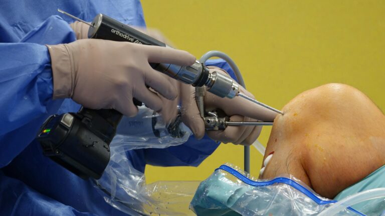
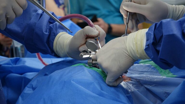
Steroid injections into the SI joint provide more aggressive pain control when conservative measures are insufficient. These injections deliver anti-inflammatory medication directly to the inflamed synovial membrane, providing up to 70% pain reduction in the following weeks. However, steroid injections are temporary—pain often returns after 6 months, and patients may need additional injections. Repeated injections carry risks including cartilage damage and systemic steroid absorption, so they’re typically limited to 3-4 per year. Surgical fusion of the SI joint is reserved for patients who fail 6+ months of conservative treatment and diagnostic injections confirming the joint as the pain source. SI joint fusion involves permanent stabilization of the joint with implants or bone graft. Recovery from fusion surgery takes 3-6 months for full healing, during which patients follow strict activity restrictions. While effective, surgery is permanent and eliminates SI joint mobility, making it a last-resort option.
Prevention Strategies and Long-Term Management
Once SI joint inflammation resolves, the risk of recurrence depends on the underlying cause. For mechanical dysfunction, maintaining strength and movement patterns learned during physical therapy prevents relapse. Regular exercise emphasizing core and hip strength, proper lifting mechanics, and awareness of posture significantly reduce recurrence risk.
For inflammatory arthritis patients, ongoing disease management with medications prescribed by a rheumatologist controls inflammation systematically, reducing SI joint flares. Pregnant women experiencing SI joint pain benefit from pelvic support belts, physical therapy, and activity modification during pregnancy. Many women experience complete pain resolution after delivery as hormonal changes reverse and the joint restabilizes, though some develop chronic pain requiring long-term management. Women who experienced SI joint pain during pregnancy have increased risk of recurrence in subsequent pregnancies, so proactive support and physical therapy in early pregnancy can prevent more severe dysfunction.
When SI Joint Inflammation Signals Systemic Disease and Requires Specialized Care
For younger patients presenting with SI joint inflammation, especially those with morning stiffness lasting more than one hour or a family history of autoimmune disease, investigation for underlying systemic arthritis is warranted. Ankylosing spondylitis, for example, is often first recognized when young patients develop SI joint pain and morning stiffness, then later develop more widespread spinal inflammation. Recognizing this early allows treatment with appropriate anti-inflammatory medications that prevent progressive spinal fusion.
Patients with SI joint inflammation that fails conservative treatment within 6-8 weeks, or whose pain is severely disabling, benefit from referral to a physiatrist (physical medicine and rehabilitation specialist) or orthopedic surgeon specializing in SI joint disorders. These specialists can perform diagnostic injections, manage advanced therapies, and determine whether surgical intervention is appropriate. Early specialist evaluation can accelerate recovery and prevent the chronic pain that develops when inflammation is inadequately managed.
Conclusion
SI joint inflammation creates pain and functional limitation affecting millions of people, but it is highly treatable. When the sacroiliac joint becomes inflamed—whether from arthritis, mechanical stress, pregnancy, or trauma—it triggers lower back and buttock pain that disrupts daily activities. The inflammation is not permanent or hopeless: approximately 95% of patients achieve significant improvement through physical therapy, activity modification, and targeted anti-inflammatory treatment within weeks to a few months.
The key to recovery is early recognition of the problem, confirmation with proper diagnosis, and consistent engagement with physical therapy to rebuild strength and movement patterns. For patients with underlying inflammatory arthritis, systemic disease management prevents recurrence. Most people never need surgery or repeated injections; disciplined conservative treatment resolves inflammation and restores function. If symptoms persist beyond 6-8 weeks despite adequate conservative care, specialist evaluation ensures correct diagnosis and guides more aggressive treatment options before chronic pain develops.