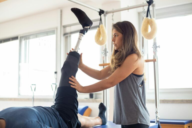
Pelvic instability occurs when the sacroiliac joints and surrounding ligaments and muscles cannot adequately stabilize the pelvis, leading to excessive joint motion or insufficient support during movement. This instability affects spine health by disrupting the load transfer between the upper body and lower extremities—the pelvis acts as a critical junction, and when it fails to distribute force properly, stress concentrates on the spine, causing pain, accelerated degeneration, and reduced mobility. The nine primary causes range from structural issues like sacroiliac joint dysfunction to hormonal changes, muscular weakness, injury, and misalignment, and understanding which factor is driving your instability is essential for effective treatment.
The connection between pelvic stability and spine health is often overlooked in mainstream discussions of back pain, yet between 15 and 30 percent of low back pain originates from sacroiliac joint pathology alone. Someone experiencing chronic lower back pain after childbirth, for instance, may assume it’s a general muscle soreness that will resolve with rest—when in fact the underlying cause could be hormonal ligament laxity, pelvic floor dysfunction, or micro-trauma from delivery, each requiring a different treatment approach. This article walks through the nine documented causes of pelvic instability, explains the biomechanical chain leading to spine stress, and clarifies why seemingly unrelated factors like pregnancy hormones or poor posture can trigger years of spinal problems if left unaddressed.
Table of Contents
- How Sacroiliac Joint Dysfunction Drives Pelvic Instability
- Hormonal Changes During Pregnancy and Ligament Laxity
- Core Muscle Weakness and Pelvic Floor Dysfunction
- Biomechanical Changes, Posture, and Center of Gravity
- Traumatic Delivery Injury and Soft Tissue Damage
- Anterior Pelvic Tilt and Load Transfer Dysfunction
- Degenerative Changes and the Long-Term Consequence of Untreated Instability
- Conclusion
How Sacroiliac Joint Dysfunction Drives Pelvic Instability
The sacroiliac joint (SIJ) is the foundation of pelvic stability—it’s a large, irregular joint at the base of the spine where the sacrum connects to the pelvis. When this joint becomes dysfunctional, it loses its ability to lock and transfer force efficiently, leaving the pelvis wobbly during everyday movements like walking, climbing stairs, or even standing. SIJ dysfunction accounts for 15 to 30 percent of low back pain cases and arises from several triggers: direct trauma (a fall onto the buttocks, a motor vehicle collision), repetitive microtrauma (years of running on hard surfaces, awkward lifting patterns), degenerative changes in the joint’s cartilage, inflammatory arthropathies like rheumatoid arthritis, or scarring after spinal surgery. The mechanism is straightforward but consequential.
A healthy sacroiliac joint has multiple strong ligaments that limit its motion to just 2-3 millimeters of movement—it’s designed to be stable, not mobile. When these ligaments are torn, stretched, or inflamed, the joint can move 10, 15, or even 20 millimeters in abnormal directions. Every step then becomes an uncontrolled experiment in load distribution; muscles downstream (in the lower back, hips, and glutes) must work harder to compensate, leading to pain, fatigue, and eventual dysfunction in those muscle groups. A runner with an old sacroiliac injury, for example, might develop hip pain, a pinched nerve, or asymmetrical knee wear because the unstable pelvis forces the leg to track incorrectly with each stride.

Hormonal Changes During Pregnancy and Ligament Laxity
Pregnancy introduces a profound hormonal shift that directly destabilizes the pelvis. The body releases relaxin and progesterone—hormones designed to soften ligaments and allow the pelvis to widen for childbirth. In practice, these hormones relax the ligaments supporting the sacroiliac joints by 25 to 40 percent, turning what was a stable, locked joint into a mobile, vulnerable one. This biochemical change begins as early as the first trimester and peaks in the third trimester and postpartum period, extending the window of vulnerability to months after delivery.
Combined with the weight of the growing fetus pulling the pelvis forward, the hormonal laxity can trigger acute sacroiliac pain during pregnancy or lay the groundwork for chronic postpartum pelvic instability. The paradox is that while relaxin prepares the pelvis for labor, it simultaneously destabilizes it for all the other functions the pelvis must perform. A pregnant woman climbing stairs, rolling over in bed, or lifting a toddler experiences pain because her pelvis is moving in ways it normally wouldn’t—yet she cannot simply “tighten” her way out of it, since the ligaments are biochemically relaxed beyond her muscular control. Some women recover their ligament tone within weeks after delivery; others struggle with sacroiliac pain for years, particularly if the ligaments are stretched excessively during a difficult labor or if postpartum rehabilitation is inadequate. This is why pelvic instability that begins in pregnancy often requires targeted intervention rather than passive waiting.
Core Muscle Weakness and Pelvic Floor Dysfunction
Beneath the bones and ligaments lies the muscular architecture of the pelvis and core—the deep abdominal muscles, the pelvic floor muscles, the multifidus, and the transverse abdominis. These muscles act as a living corset, contracting before movement to pre-stabilize the sacroiliac joint and maintain pressure within the abdominal cavity. When these muscles are weak, inhibited, or misfiring, the pelvis loses dynamic stability. Research shows that women have inherently weaker pelvic floor muscles than men, increasing baseline susceptibility to low back pain; pregnancy then exacerbates this by stretching the core muscles and temporarily reducing their functional capacity as the abdominal wall expands to accommodate the fetus.
Pelvic floor dysfunction—present in 52 percent of women experiencing pregnancy-related low back and pelvic pain—involves more than simple weakness; it includes altered firing patterns where muscles contract at the wrong time or with insufficient force. A woman with pelvic floor dysfunction might feel heaviness or pressure, inability to control urinary leakage, or pain during intercourse, and these symptoms correlate strongly with sacroiliac instability because the pelvic floor is one of the primary stabilizers. Physical therapy addressing these muscles with specific exercises and biofeedback can restore the neural activation patterns, allowing the pelvis to re-stabilize even if hormonal laxity or weakness persists. However, if pelvic floor weakness is not specifically rehabilitated—and instead the focus is only on general core strengthening—improvement may be slow or incomplete, leading to frustration for patients expecting recovery.

Biomechanical Changes, Posture, and Center of Gravity
The pelvis does not exist in isolation; it is part of a kinetic chain where posture, weight distribution, and alignment directly affect how load passes through the sacroiliac joint. During pregnancy and with advancing age, the body’s center of gravity shifts forward as weight increases, forcing the pelvis to anteriorly rotate—tilt forward—to maintain balance. This forward pelvic tilt increases lumbar lordosis (the inward curve of the lower back), concentrating compressive and shear forces on the sacroiliac joint rather than distributing them evenly. Additionally, muscles in the hip flexors tighten with prolonged sitting or poor postural habits, pulling the pelvis further into anterior tilt and overloading the posterior ligaments of the sacroiliac joint. The biomechanical consequences are measurable and progressive.
A person with anterior pelvic tilt experiences increased load on the posterior sacroiliac joint ligaments—the very tissues that are already stressed or strained by hormonal laxity or prior injury. Over weeks and months, this chronic overload causes inflammation, pain with certain movements, and eventual degenerative changes in the joint. Conversely, posterior pelvic tilt—tilting the pelvis backward—reduces sacroiliac load but often reflects muscular inhibition or postural collapse, creating its own problems. The optimal posture is neutral—a slight anterior tilt that allows the core muscles to engage and the sacroiliac ligaments to lock. Someone with chronic poor posture from desk work, for instance, may develop sacroiliac instability not from a single injury but from years of anterior pelvic tilt and associated ligament stress, illustrating how biomechanical factors act silently over time.
Traumatic Delivery Injury and Soft Tissue Damage
Childbirth is a profound physical event, and complicated labor—involving a contracted pelvis, a large baby, shoulder dystocia (the baby’s shoulder catches on the pubic bone), or a prolonged second stage of labor—creates significant mechanical stress on the pelvis. The forces exerted during pushing and the passage of the fetus can stretch, tear, or irritate the sacroiliac ligaments, the pelvic floor muscles, and associated fascia. In some cases, fractures of the pubic symphysis or sacroiliac joint occur, though these are rare in developed countries. More commonly, the soft tissue damage from delivery is not immediately obvious—pain is attributed to general postpartum soreness—but the underlying ligament or muscular injury sets the stage for chronic instability if not specifically rehabilitated.
The distinction between normal postpartum recovery and pathological instability is timing and severity. Most women experience some pelvic pain in the immediate postpartum weeks as tissues heal; this typically resolves within 3 to 6 months with gradual return to activity. However, women with significant delivery-related soft tissue injury—particularly those with unassisted traumatic delivery, multiple deliveries in close succession, or obesity—face higher risk of persistent sacroiliac dysfunction if rehabilitation is not targeted and progressive. A woman whose instability stems primarily from delivery trauma often benefits from early, specific pelvic physical therapy in the weeks following birth, whereas waiting 6 months for pain to spontaneously resolve may allow compensatory movement patterns to entrench, making later recovery slower and harder. This is a critical limitation of the “wait and see” approach to postpartum pelvic pain.

Anterior Pelvic Tilt and Load Transfer Dysfunction
Anterior pelvic tilt—the forward rotation of the pelvis relative to the spine and femurs—deserves specific attention because it is both common and modifiable, and because it directly impairs load transfer efficiency. When the pelvis tilts anteriorly, the angle of the sacroiliac joint changes, shifting load distribution away from the ideal alignment. The posterior sacroiliac ligaments, which are strongest when the joint is locked in a specific position, become slack or overloaded depending on the degree of tilt. Simultaneously, the lumbar spine is forced into excessive extension (backward bending), concentrating stress on the small facet joints of the lower back rather than distributing it through the large sacroiliac joint and into the legs.
The practical consequence is that someone with anterior pelvic tilt experiences pain not just at the sacroiliac joint but up and down the spine—a classic pattern is lower back pain that worsens with walking or standing. Interestingly, many people with anterior pelvic tilt are unaware of their postural deviation, attributing their pain to general “back problems” rather than recognizing that realigning the pelvis could resolve or significantly improve their symptoms. Postural correction through awareness, specific muscle retraining (hip flexor stretching, glute activation), and core stabilization can normalize load transfer, but it requires consistency and often professional guidance to retrain decades of habitual posture. Simply doing generic core exercises like crunches or planks often worsens anterior pelvic tilt if the hip flexors are not simultaneously lengthened.
Degenerative Changes and the Long-Term Consequence of Untreated Instability
When pelvic instability persists unchecked for months or years, the excessive, abnormal motion of the sacroiliac joint accelerates degenerative changes. The cartilage surface of the joint experiences uneven compression and shear stress; the bone beneath can develop sclerosis (hardening) and osteophytes (bone spurs); and the joint capsule becomes inflamed and thickened. This degenerative process is not unique to the sacroiliac joint—it mirrors osteoarthritis in any joint exposed to chronic mechanical stress—but it is particularly significant because the sacroiliac joint, unlike the hip or knee, has limited treatment options once significant degeneration occurs. Early intervention to restore stability slows or halts degeneration; late intervention, when cartilage loss is already advanced, may require more invasive treatment such as sacroiliac joint fusion surgery.
The trajectory from acute instability to chronic degeneration is often preventable. A person with a recent sacroiliac injury who receives appropriate physical therapy and activity modification can stabilize the joint and prevent degeneration. Conversely, someone who ignores persistent sacroiliac pain for 5 or 10 years—assuming it will eventually resolve or accepting it as an inevitable part of aging—risks developing degenerative joint disease that reduces mobility and quality of life. This underscores the importance of early recognition and treatment of pelvic instability, particularly after pregnancy or injury when the instability is most treatable. The paradox is that early-stage instability often feels manageable or attributable to other causes, reducing the urgency to seek care, yet this delay is when the most effective interventions are possible.
Conclusion
Pelvic instability arises from multiple, often overlapping causes—sacroiliac joint dysfunction, hormonal changes, muscular weakness, biomechanical misalignment, delivery trauma, and degeneration—each contributing to the loss of the pelvis’s critical role in distributing load between the spine and legs. Understanding which cause or combination of causes is driving your instability is the first step toward targeted, effective treatment. An athlete with SIJ dysfunction from repetitive impact requires a different approach than a postpartum woman with hormonal laxity and pelvic floor dysfunction, yet both conditions are labeled “pelvic instability” and risk being treated generically with standard core exercises that may not address the underlying pathology.
If you experience persistent lower back or pelvic pain, particularly pain that worsens with specific movements, after pregnancy, or following an injury, evaluation by a healthcare provider trained in sacroiliac joint dysfunction and pelvic physical therapy is essential. Early diagnosis and treatment—including targeted physical therapy, postural correction, and activity modification—can prevent the progression to chronic pain and degenerative joint changes. Conversely, delaying care in hopes that instability will self-resolve increases the risk of compensatory dysfunction in the hips, knees, and spine, and prolongs suffering that could otherwise be addressed in weeks to months of focused rehabilitation.