Six physical therapy movements commonly used to strengthen the sacroiliac joint include the bridge, the clamshell, the bird-dog, transverse abdominis activation, piriformis stretching, and supine SI joint reset techniques. These exercises target the muscles surrounding the SI joint — where the sacrum meets the ilium in the pelvis — and are frequently prescribed by physical therapists because they address both stability and mobility without placing excessive load on the joint itself. For someone recovering from SI joint dysfunction, a structured routine incorporating these movements three to five times per week can gradually reduce pain and restore functional movement, though the specific combination and intensity should always be tailored by a qualified therapist.
This matters more than many people realize for aging populations, including those navigating cognitive decline. Chronic pain from SI joint instability can reduce mobility, disrupt sleep, and accelerate the kind of physical deconditioning that worsens outcomes in dementia care. A person with early-stage Alzheimer’s who also has untreated SI joint pain, for instance, may stop walking regularly — and that lost activity compounds both physical and cognitive decline. This article walks through each of the six movements in detail, explains why they work, discusses when they may not be appropriate, and addresses how SI joint health connects to broader well-being in older adults.
Table of Contents
- Why Are These Six Physical Therapy Movements Recommended for the SI Joint?
- Bridging and Clamshells — Building the Foundation of Pelvic Stability
- The Bird-Dog and Core Activation for Joint Protection
- How to Perform Transverse Abdominis Activation and Piriformis Stretching Safely
- When SI Joint Exercises Can Backfire — Warnings and Limitations
- The Supine SI Joint Reset and When to Use It
- SI Joint Health as Part of Whole-Person Care in Aging and Dementia
- Conclusion
- Frequently Asked Questions
Why Are These Six Physical Therapy Movements Recommended for the SI Joint?
The SI joint is inherently a low-motion joint, designed more for load transfer than for range of movement. It relies heavily on the muscles and ligaments around it for stability. When those structures weaken — from aging, prolonged sitting, injury, or conditions like arthritis — the joint can become hypermobile or misaligned, producing pain that radiates through the low back, buttock, and sometimes down the leg. Physical therapists target this problem not by mobilizing the joint itself but by strengthening the muscular scaffolding that holds it in place. The six movements listed above were not chosen arbitrarily. Each one addresses a specific muscle group or movement pattern that contributes to SI joint stability.
The bridge targets the gluteus maximus and hamstrings, which support the posterior pelvis. The clamshell isolates the gluteus medius, a muscle responsible for lateral hip stability. The bird-dog trains coordinated core activation across the trunk and pelvis. Transverse abdominis work engages the deepest layer of abdominal muscle, which acts like a natural corset around the pelvis. Piriformis stretching addresses a muscle that, when tight, can pull the sacrum out of alignment. And supine SI reset techniques use gentle isometric contractions to restore symmetry when the joint feels “stuck.” Compared to generalized core exercise programs, this targeted combination addresses the specific biomechanical demands of the SI joint rather than broadly strengthening the trunk.

Bridging and Clamshells — Building the Foundation of Pelvic Stability
The bridge is often the first exercise prescribed for SI joint dysfunction because it is low-impact, easily modified, and highly effective at activating the gluteal muscles. To perform it, a person lies on their back with knees bent and feet flat on the floor, then lifts the hips toward the ceiling while squeezing the glutes. The movement should be slow and controlled, with a brief hold at the top. For someone who finds the standard bridge too demanding, a therapist might start with a partial bridge — lifting the hips only a few inches — or place a pillow between the knees to encourage better alignment. The clamshell works differently.
Lying on one side with knees bent at roughly 45 degrees, the person opens the top knee while keeping the feet together, like a clamshell opening. This isolates the gluteus medius, a muscle that many people neglect entirely but that plays a critical role in preventing the pelvis from dropping or shifting laterally during walking. Weakness in the gluteus medius is one of the most common contributors to SI joint dysfunction, particularly in people who sit for long periods. However, if someone has significant hip osteoarthritis or a labral tear, the clamshell can sometimes aggravate hip pain rather than help. In these cases, a therapist may substitute a side-lying hip abduction with the leg straighter, or use a resistance band at a lower intensity. The point is that no exercise is universally safe — context matters, and a movement that helps one person’s SI joint can irritate another person’s hip.
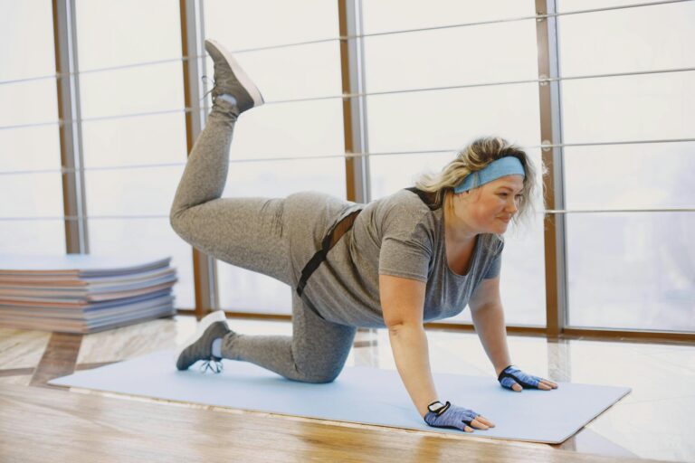
The Bird-Dog and Core Activation for Joint Protection
The bird-dog is a deceptively simple exercise that challenges balance, coordination, and core stability simultaneously. Starting on hands and knees, the person extends the opposite arm and leg while keeping the trunk completely still. The goal is not to lift as high as possible but to maintain a neutral spine throughout the movement. This trains the body to stabilize the pelvis under dynamic conditions — exactly the kind of demand that daily activities like walking, bending, and reaching place on the SI joint.
What makes the bird-dog particularly valuable for older adults is that it also trains proprioception, the body’s sense of where it is in space. Proprioceptive deficits are a known fall risk factor in aging populations and are often more pronounced in people with cognitive impairment. A 2019 review in the Journal of Geriatric Physical Therapy noted that exercises combining balance and strength components, like the bird-dog, had measurable effects on fall reduction in older adults, though specific data on SI joint outcomes in dementia populations remains limited. For someone who cannot safely get onto hands and knees — due to wrist arthritis, knee pain, or balance concerns — the bird-dog can be modified to a standing position using a countertop for support, or performed from a seated position on a stability ball. A physical therapist working with a dementia patient might also simplify the movement to single-limb extensions rather than opposite arm-and-leg combinations, reducing the cognitive load of the task while still providing the stabilization benefit.

How to Perform Transverse Abdominis Activation and Piriformis Stretching Safely
Transverse abdominis (TA) activation is not a flashy exercise. It involves lying on the back with knees bent and gently drawing the lower abdomen inward, as if pulling the belly button toward the spine. There is no visible movement. The contraction is subtle, internal, and easy to do incorrectly. Many people compensate by holding their breath, bracing the rectus abdominis (the “six-pack” muscle), or tilting the pelvis — all of which defeat the purpose. A skilled physical therapist will often use tactile or verbal cues, and sometimes real-time ultrasound imaging, to ensure the correct muscle is firing. The tradeoff with TA activation is that it requires a level of body awareness and concentration that can be difficult for people with moderate to advanced cognitive impairment.
In early-stage dementia, most individuals can learn the activation with patient instruction and repetition. In later stages, a therapist may shift to exercises that recruit the TA indirectly — like modified bridging with a focus on slow, controlled breathing — rather than asking for isolated activation that the patient cannot reliably perform. Piriformis stretching, by contrast, is more intuitive. The classic version involves lying on the back, crossing one ankle over the opposite knee, and pulling the uncrossed leg toward the chest. This produces a stretch deep in the buttock where the piriformis muscle runs from the sacrum to the femur. When the piriformis is chronically tight, it can compress the sciatic nerve and pull the sacrum into a rotated position, destabilizing the SI joint. Regular stretching — held for 30 to 60 seconds, repeated two or three times per side — can relieve this tension. However, people with hip replacements should avoid this position unless their surgeon has specifically cleared it, as the cross-legged posture can risk dislocation depending on the surgical approach used.
When SI Joint Exercises Can Backfire — Warnings and Limitations
Not every case of SI joint pain responds to strengthening exercises, and there are scenarios where performing them without proper evaluation can make things worse. The most important distinction a physical therapist makes is between SI joint hypermobility and SI joint hypomobility. Hypermobility means the joint moves too much and needs stabilization — the exercises described in this article are designed for this situation. Hypomobility means the joint is locked or restricted, and attempting to stabilize it further can increase stiffness and pain. A locked SI joint typically requires manual therapy or mobilization techniques first, followed by stabilization work once normal movement is restored. Another limitation involves inflammatory conditions.
Ankylosing spondylitis, for example, is an autoimmune condition that commonly affects the SI joints and causes progressive fusion. Exercising through an active inflammatory flare in this context is counterproductive and can increase damage. Similarly, SI joint pain that does not improve after four to six weeks of consistent physical therapy may indicate an underlying issue — such as a stress fracture, infection, or tumor — that requires imaging and medical evaluation rather than continued exercise. For caregivers supporting someone with dementia who also has SI joint pain, there is an additional concern: the person may not be able to accurately report whether an exercise is helping or hurting. Pain assessment in cognitively impaired individuals often requires observation of behavioral cues — facial grimacing, resistance to movement, changes in mood or agitation — rather than relying on verbal self-report. If a caregiver notices increased agitation or resistance during or after exercises, it is worth pausing the routine and consulting the prescribing therapist.

The Supine SI Joint Reset and When to Use It
The supine SI joint reset is a gentle technique that physical therapists sometimes teach patients for self-management between sessions. One common version involves lying on the back, bending one knee to the chest, and pressing the knee into the hands with moderate force — an isometric contraction held for five to ten seconds — then releasing.
The same is done on the opposite side, followed by pressing both knees outward against the hands and then inward against a ball or pillow placed between the knees. The series of isometric contractions helps recalibrate the muscular tension around the SI joint and can provide temporary relief when the joint feels asymmetric or “off.” This technique is not a substitute for strengthening work, and it does not fix the underlying instability that causes SI joint dysfunction. Think of it as a reset button rather than a repair — useful for managing flare-ups, particularly after prolonged sitting or an awkward movement, but most effective when combined with the other five exercises described above.
SI Joint Health as Part of Whole-Person Care in Aging and Dementia
There is growing recognition in geriatric medicine that musculoskeletal pain and cognitive decline interact in ways that are difficult to untangle. Chronic pain increases cortisol levels, disrupts sleep architecture, reduces physical activity, and contributes to social withdrawal — all of which are independently associated with faster cognitive decline. Addressing SI joint dysfunction in an older adult is not just about reducing back pain; it is about preserving the physical capacity that supports independence, engagement, and quality of life.
Physical therapy programs that incorporate SI joint work alongside balance training, walking endurance, and functional movement patterns offer the most comprehensive benefit. As research into the overlap between chronic pain management and dementia care continues to develop, there is reason to expect that integrated approaches — where physical therapists, neurologists, and primary care providers coordinate treatment — will become the standard rather than the exception. For now, the practical step is straightforward: if an older adult or someone in your care has persistent low back or pelvic pain, a physical therapy evaluation that specifically assesses the SI joint is a reasonable and low-risk starting point.
Conclusion
The six physical therapy movements discussed here — bridges, clamshells, bird-dogs, transverse abdominis activation, piriformis stretching, and supine SI joint resets — represent a well-established approach to stabilizing the sacroiliac joint. Each targets a different component of the muscular system that supports pelvic alignment, and together they address the most common causes of SI joint instability. They are low-impact, adaptable, and appropriate for most older adults when guided by a qualified physical therapist.
For those managing dementia alongside chronic pain, the stakes are higher and the execution more nuanced. Exercises may need to be simplified, pain must be assessed through observation as much as self-report, and the overall goal should be preserving mobility and comfort rather than achieving athletic performance. A physical therapist experienced in working with cognitively impaired patients can make meaningful adjustments. The key takeaway is that SI joint pain is treatable, that targeted exercises work for many people, and that letting the pain go unaddressed carries its own set of consequences — reduced movement, worse sleep, and a steeper decline in the capacities that matter most.
Frequently Asked Questions
How long does it take for SI joint exercises to reduce pain?
Most physical therapists advise giving a consistent exercise program four to six weeks before evaluating its effectiveness. Some people notice improvement within two weeks, while others require several months. If there is no improvement after six weeks of consistent work, further evaluation is warranted.
Can SI joint exercises be done at home without a physical therapist?
After an initial evaluation and instruction, most of these exercises can be performed at home. However, the first few sessions should be supervised to ensure correct form, especially for transverse abdominis activation, which is frequently performed incorrectly without professional guidance.
Is walking good or bad for SI joint pain?
Walking is generally beneficial for SI joint health because it promotes gentle, rhythmic movement through the pelvis. However, walking on uneven terrain or for prolonged distances can aggravate an unstable SI joint. Starting with short, flat-surface walks and gradually increasing duration is the safest approach.
Should I use a sacroiliac belt along with these exercises?
SI belts can provide temporary external stability and are sometimes recommended during the early phase of rehabilitation when the joint is most irritable. They are not a long-term solution. The goal of the exercises is to build internal stability so that a belt becomes unnecessary.
Are these exercises safe for someone with osteoporosis?
Most of these exercises are low-impact and generally safe for people with osteoporosis, but the bird-dog and bridge should be performed carefully to avoid excessive spinal extension. A physical therapist familiar with osteoporosis precautions can modify the program appropriately.