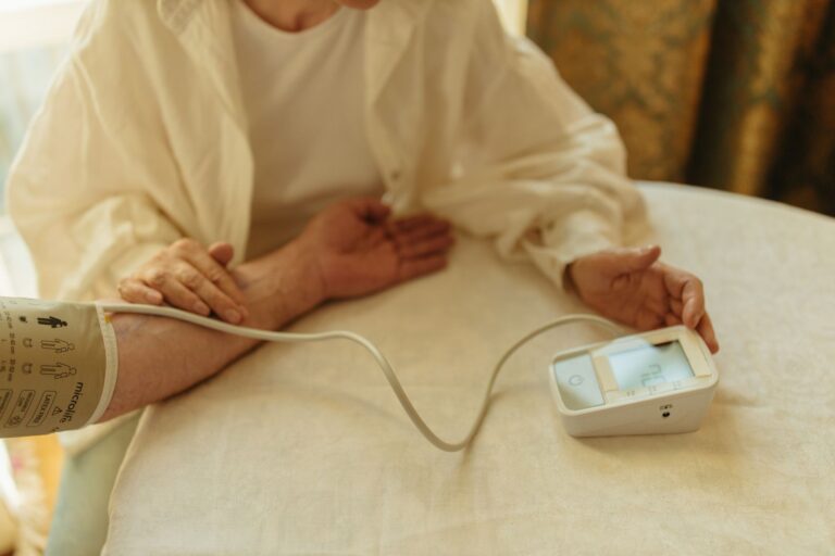
Researchers measuring exercise’s effects on Alzheimer’s disease track far more than whether someone remembers where they parked. In clinical trials, scientists measure cognitive scores from standardized tests, blood and brain biomarkers that indicate disease progression, physical fitness metrics, imaging changes in brain structure, and how well people manage daily tasks. These multiple measures are necessary because Alzheimer’s affects the brain in complex ways—exercise might slow cognitive decline in one person while improving their balance and mood in another, and researchers need tools to capture all these changes. A real example: the Aerobic Exercise and Cognitive Decline (ACED) study tracked people with mild cognitive impairment using the Montreal Cognitive Assessment (MoCA) to measure thinking skills, blood tests for inflammatory markers, MRI brain scans to look for volume changes in the hippocampus, and gait speed tests to assess how exercise affected walking.
No single measurement told the whole story. Some participants showed no change in memory scores but significant improvements in processing speed or how long they could exercise before tiring. Why multiple measurements? Alzheimer’s disease involves amyloid plaques, tau tangles, inflammation, vascular damage, and neurodegeneration all happening at once. A trial measuring only cognition might miss that exercise reduced a person’s fall risk by 40% or improved their sleep patterns. Researchers also recognize that cognitive tests alone don’t capture quality of life—someone might score slightly lower on a memory test but regain the independence to cook meals or manage their medications.
Table of Contents
- What Cognitive Tests Measure in Alzheimer’s Exercise Trials
- Biomarkers—Measuring the Biology Behind the Symptoms
- Brain Imaging and Structural Changes
- Physical Fitness and Functional Capacity Testing
- Daily Living Skills and Instrumental ADL
- Mood, Sleep, and Behavioral Measures
- Specific Biomarkers and Inflammatory Markers
What Cognitive Tests Measure in Alzheimer’s Exercise Trials
The Mini-Cog, Montreal cognitive Assessment (MoCA), and Alzheimer’s Disease Assessment Scale–cognitive subscale (ADAS-cog) are the workhorses of exercise trials. The ADAS-cog, used in major pharmaceutical trials, includes word recall, naming, following commands, and attention tasks, with scores ranging from 0 to 70—higher scores indicate worse cognition. A typical trial might look for a slowing of decline, where untreated patients drop 2–3 points per year on the ADAS-cog, but exercise groups decline only 1 point yearly. These tests measure different cognitive domains because Alzheimer’s doesn’t erode the brain uniformly. Executive function (planning, problem-solving) declines differently than episodic memory (remembering specific events) or processing speed (how fast the brain responds). Some exercise trials show stronger benefits for processing speed and attention than for memory.
For instance, a trial of aerobic exercise at a senior center might find participants faster at completing a timed number-cancellation task but show only modest gains in delayed verbal recall. Researchers report both findings because they reveal which cognitive systems respond best to physical activity. One limitation: cognitive tests are influenced by education level, cultural background, and language. A person who speaks English as a second language might score lower on the MMSE not because of Alzheimer’s progression but because the test uses English-language word associations. This means trials must carefully match study groups and sometimes adjust scores, yet subtle biases can remain. Researchers also note that coaching matters—someone who has taken a cognitive test twice may perform better the third time simply from familiarity, not from actual improvement.
Biomarkers—Measuring the Biology Behind the Symptoms
Blood biomarkers have transformed Alzheimer’s research over the past five years. Tests now measure phosphorylated tau (p-tau), amyloid-beta ratios, neurofilament light chain (NfL), and glial fibrillary acidic protein (GFAP) in a small blood draw. These proteins leak from damaged neurons and accumulate in the bloodstream, making them windows into brain pathology without requiring a spinal tap or brain biopsy. In exercise trials, researchers compare biomarker levels between the exercise group and the control group over 6, 12, or 24 months. One study of resistance training in people with cognitive impairment measured blood amyloid-beta-42 and found that the exercise group had lower levels of a specific amyloid fragment associated with plaque formation, while the control group showed the typical decline. Another trial combining aerobic exercise with cognitive training measured p-tau181 and found slower biomarker progression in the active group.
However, biomarker changes don’t always match cognitive outcomes—a person might show improved blood biomarkers but no change in memory scores, or vice versa. This disconnect is why researchers don’t rely on biomarkers alone. Cerebrospinal fluid (CSF) biomarkers, obtained from a lumbar puncture, offer even more direct evidence of brain changes but are invasive and limit study enrollment. Most large exercise trials now use blood biomarkers instead, which are far easier to collect repeatedly. A caveat: the commercial blood tests are still being standardized, and different labs may report slightly different values. This is improving as the tests become more established, but it means early trial results should be interpreted cautiously until the assays are locked down.
Brain Imaging and Structural Changes
MRI (magnetic resonance imaging) allows researchers to measure brain volume, particularly in the hippocampus, the seahorse-shaped region critical for memory that often shrinks in Alzheimer’s disease. A typical progression involves 2–3% annual hippocampal volume loss in untreated older adults with mild cognitive impairment. Exercise trials test whether this loss slows or halts. One notable trial found that older adults who walked briskly four times weekly for one year showed a small increase in hippocampal volume, while sedentary controls lost volume over the same period. PET imaging (positron emission tomography) is more expensive and less commonly used in exercise trials but offers a direct view of amyloid and tau accumulation in the brain. A PET scan shows regions “lighting up” where pathological proteins cluster.
Some exercise trials use PET to see if exercise slows the spread of these deposits, but most rely on the less costly MRI, which still detects changes in brain structure, white matter integrity, and regional atrophy. Functional MRI (fMRI) measures blood flow during cognitive tasks, revealing which brain regions are “working” and whether exercise changes the brain’s activity patterns. A limitation is that fMRI signals are small and noisy—changes that look impressive in a single brain scan may not replicate across many participants, so meta-analyses (combining multiple small trials) are essential for reliable conclusions. DTI (diffusion tensor imaging) measures the integrity of white matter tracts, the wiring connecting brain regions. Alzheimer’s disease degrades these pathways, and some researchers hypothesize that aerobic exercise strengthens them. Detecting these changes requires highly sensitive imaging and large sample sizes. For this reason, most trials use standard structural MRI, which is faster and more accessible across different hospital scanners.
Physical Fitness and Functional Capacity Testing
Researchers measure cardiorespiratory fitness using a treadmill test or six-minute walk test, where participants walk as far as possible in six minutes on a flat hallway. This predicts mortality and disability risk independent of cognitive status. An exercise trial might show that a group doing 150 minutes weekly of moderate aerobic activity increases their six-minute walk distance by 50–100 meters over a year, while a sedentary control group shows no change or slight decline. Leg strength, measured using a leg press machine or chair stand test (counting how many times someone can stand and sit from a chair without using their arms in 30 seconds), directly predicts the ability to climb stairs, get out of bed, and maintain independence. Balance tests like the Timed Up and Go (rising from a chair, walking 10 feet, turning around, and returning) capture fall risk. A person with Alzheimer’s and poor balance scores (>12 seconds on the Timed Up and Go) has a significantly higher fall and fracture risk, making this measurement critical.
Exercise trials often show improvements in Timed Up and Go scores among participants doing resistance or balance training, even when cognition doesn’t improve. Hand grip strength correlates with overall muscle mass and function. It’s quick to measure (using a hand dynamometer) and predicts mortality and functional decline. Researchers include it as a marker of whether exercise is building lean muscle mass or merely improving cardiovascular fitness. A tradeoff: measuring many fitness domains gives a complete picture but requires more testing time per participant and can fatigue people, especially those with advanced Alzheimer’s who tire easily. Trials often select 3–5 key fitness measures rather than assessing everything possible.
Daily Living Skills and Instrumental ADL
The Activities of Daily Living (ADL) scale measures ability to dress, bathe, use the toilet, eat, and walk independently. The Instrumental ADL (IADL) scale assesses higher-level tasks: preparing meals, managing finances, handling medications, housekeeping, laundry, using a phone, and managing transportation. Alzheimer’s typically affects IADL before ADL—someone might forget to pay bills or take their pills while still able to shower themselves. Exercise trials include IADL measures because maintaining independence in these tasks is often what keeps people living at home rather than in assisted care. Caregiver-reported IADL change correlates better with quality of life than cognitive scores do.
A person might score unchanged on a memory test but become unable to safely use a stove, requiring the caregiver to take over meal preparation entirely. Conversely, exercise that improves attention and motor coordination might help someone remember their medication schedule and prepare simple meals, reducing caregiver burden. A 12-week exercise trial might measure IADL at baseline, 12 weeks, and 24 weeks, with the primary question being whether the exercise group declines more slowly than controls. One limitation: caregivers report IADL status, and caregivers’ own stress, depression, or time availability influence how accurately they observe and report changes. A caregiver working full-time might not notice that their spouse (with Alzheimer’s) no longer initiates household tasks but could do them if asked, leading to underestimation of remaining ability. Trials often employ standardized questionnaires and train raters to improve consistency.
Mood, Sleep, and Behavioral Measures
Depression and apathy are common in Alzheimer’s and are measurable using the Geriatric Depression Scale (GDS) and other instruments. Exercise often improves mood, so many trials include depression scores as secondary outcomes. Sleep quality, measured via questionnaires or actigraphy (a wearable that detects sleep and wake patterns), frequently improves with exercise.
A trial might find that participants in an aerobic exercise program report fewer nighttime awakenings and longer consolidated sleep, which often leads to better daytime function and less agitation. Agitation and behavioral disturbances, measured on scales like the Neuropsychiatric Inventory (NPI), may improve or sometimes worsen with exercise depending on exercise timing (overexertion in the evening might increase nighttime agitation) and individual tolerance. Anxiety levels, tracked via standardized questionnaires, sometimes decrease as people gain confidence and structure from regular exercise routines. Researchers document these behavioral changes because they have real impacts: someone who is less agitated is easier to care for, has better sleep, and may have fewer psychotropic medication needs.
Specific Biomarkers and Inflammatory Markers
Beyond amyloid and tau, researchers measure inflammatory proteins like IL-6 (interleukin-6), TNF-alpha, and C-reactive protein (CRP). Chronic inflammation is implicated in Alzheimer’s progression, and exercise reduces systemic inflammation in many populations. A trial comparing a walking group to a control group might measure serum IL-6 levels at baseline and at the end of the study, finding that the walking group’s IL-6 levels dropped 15–20% while the control group’s remained stable or rose slightly. This suggests that exercise has an anti-inflammatory effect that could slow cognitive decline.
One exercise trial in people with mild cognitive impairment measured both blood IL-6 and cognitive scores, finding a modest correlation: participants with the largest reductions in IL-6 tended to show slower cognitive decline. However, this association is far from universal, and another trial might find no such relationship. The variability reflects that each person’s brain is different—some people’s cognitive problems stem heavily from inflammation, while others have more amyloid pathology or vascular damage. This is why researchers measure multiple biomarkers and why single studies never settle the question of exercise’s mechanism.
- —