Doctors identify seven primary causes of sacroiliac joint dysfunction in clinical practice: trauma or repetitive injury, pregnancy-related hormonal changes, osteoarthritis and degenerative joint disease, post-surgical instability, inflammatory arthropathies, biomechanical abnormalities including leg length discrepancy, and obesity combined with occupational or athletic overuse. These conditions account for the majority of sacroiliac joint (SIJ) pain cases seen in musculoskeletal medicine practices, physical therapy clinics, and orthopedic settings.
A 45-year-old construction worker presenting with low back pain radiating to one side might have developed SIJ dysfunction from years of repetitive bending and lifting, while a postpartum woman experiencing similar pain may have hormonal joint laxity as the underlying cause. Despite affecting 15-30% of chronic low back pain patients, SIJ dysfunction remains underdiagnosed because many clinicians focus exclusively on lumbar spine pathology. This article explores each of the seven clinical causes doctors see regularly, the mechanisms behind each, relevant statistics from medical literature, and how proper diagnosis depends on recognizing which specific cause is driving a patient’s symptoms.
Table of Contents
- What Role Does Direct Injury Play in Sacroiliac Joint Problems?
- How Does Pregnancy Change the Sacroiliac Joint and Why Is Recovery Important?
- Why Is Osteoarthritis the Most Common Cause Doctors Encounter?
- How Does Spine Surgery Sometimes Create Sacroiliac Joint Problems?
- What Do Inflammatory Arthropathies Like Ankylosing Spondylitis Cause in the Sacroiliac Joint?
- How Do Biomechanical Problems and Weight Imbalance Load the Sacroiliac Joint?
- How Do Doctors Diagnose Which of These Seven Causes Is Present?
- Conclusion
What Role Does Direct Injury Play in Sacroiliac Joint Problems?
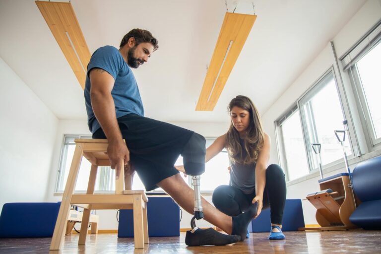
Trauma and repetitive microtrauma represent one of the most straightforward causes of SIJ dysfunction. A direct fall onto the buttock, a motor vehicle accident with side-impact forces, or a sports injury involving sudden twisting can acutely damage sacroiliac joint ligaments and capsular structures. However, many SIJ problems develop more insidiously through occupational or athletic repetitive stress—a roofer climbing and descending ladders hundreds of times monthly, a runner logging high mileage with poor running mechanics, or a warehouse worker repeatedly lifting boxes from low shelves.
These repetitive microtrauma cases often involve a specific inciting event that patients can pinpoint, such as a lifting injury during a single heavy load, after which symptoms gradually worsen over weeks. The distinction between acute trauma and repetitive microtrauma matters clinically because it affects treatment expectations. Acute traumatic injuries may require more aggressive initial pain management and shorter periods of immobilization through pelvic bracing, while repetitive strain cases typically respond better to activity modification, biomechanical correction, and progressive stabilization exercises. However, even identical-appearing injuries can progress differently depending on whether the patient’s ligaments were predisposed to injury through prior joint laxity, hormonal factors, or genetic connective tissue differences.

How Does Pregnancy Change the Sacroiliac Joint and Why Is Recovery Important?
Pregnancy creates a unique and temporary vulnerability in the sacroiliac joints through hormonal mechanisms. During pregnancy, the body releases relaxin and other hormones that increase ligament laxity to prepare for childbirth, but this same hormonal effect destabilizes the sacroiliac joints. The additional weight of the gravid uterus, combined with postural changes as the center of gravity shifts forward, concentrates mechanical stress on already-loosened SIJ ligaments. Studies show pregnant women experience significantly higher rates of SIJ dysfunction than age-matched non-pregnant women, and symptoms frequently persist into the postpartum period if not specifically addressed.
Treatment during and after pregnancy has evolved substantially. Pelvic girdle stabilization using a specifically designed pelvic support belt—worn snugly around the pelvis below the level of the sacroiliac joints—provides external stability that compensates for hormonal laxity. Postpartum pelvic belts have demonstrated clinical benefit when used in the first weeks and months after delivery while hormonal joint mobility gradually normalizes. However, if SIJ pain is ignored during pregnancy or if a woman returns immediately to high-impact activities postpartum before adequate stabilization has returned, symptoms can become chronic, persisting for years after delivery. Patient education about gradual return to exercise and appropriate pelvic floor rehabilitation represents a critical but often-overlooked component of postpartum care.
Why Is Osteoarthritis the Most Common Cause Doctors Encounter?
Osteoarthritis and degenerative changes of the sacroiliac joint represent the most frequent cause of SIJ dysfunction identified in clinical practice. As with any synovial joint, the cartilage covering the sacroiliac articular surfaces can deteriorate with age, wear, and prior injury. This degenerative process creates structural instability because the joint loses its smooth, congruent surfaces and develops irregular osteophytes (bone spurs) and cartilage loss. Once degeneration begins, the joint compensates by increasing its reliance on ligaments and muscles for stability, yet these supporting structures may themselves be atrophied or weakened, creating a vicious cycle of increasing pain and dysfunction.
The prevalence of SIJ osteoarthritis increases steadily with age, particularly after age 50, making it a leading cause in older populations. A 62-year-old woman with decades of chronic low back pain from a prior lumbar fusion may develop severe SIJ osteoarthritis without realizing the pain has shifted from the lumbar spine to the sacroiliac joint. Importantly, some patients with extensive degenerative changes visible on imaging experience no pain, while others with minimal radiographic findings suffer significantly, highlighting that osteoarthritis alone does not explain all cases. This disconnect underscores why imaging must be paired with clinical examination—multiple positive SIJ provocation tests indicate that degeneration is symptomatic rather than simply incidental.

How Does Spine Surgery Sometimes Create Sacroiliac Joint Problems?
Post-surgical instability represents a well-recognized iatrogenic cause of SIJ dysfunction. Lumbar fusion surgery, particularly fusion at L5-S1, fundamentally alters the biomechanics of the lumbosacral region. By eliminating motion between two vertebrae, fusion surgery increases mechanical stress on all adjacent joints, including the sacroiliac joints. This biomechanical shift concentrates load unevenly across the sacroiliac articulation, accelerating degeneration and increasing ligament strain. The longer the fusion construct (multi-level fusions increase the effect), the greater the stress transferred to the SIJ.
Up to 40-63% of patients with failed back surgery syndrome—persistent pain after one or more spinal surgeries—have SIJ pain as the pain source rather than problems at the surgical site. Recognition of post-surgical SIJ dysfunction has grown as more patients undergo fusion procedures and subsequently develop new or worsening symptoms years later. A 55-year-old patient who had successful pain relief from L4-L5 fusion may develop sacroiliac pain five years later from exactly this mechanism. Treatment in post-surgical patients requires different strategies than primary SIJ dysfunction because surgical hardware limits manipulation options and may preclude certain injection approaches. However, non-operative management with targeted stabilization exercises, activity modification, and radiofrequency ablation of intra-articular nerves often provides adequate relief without reoperation. When conservative care fails, sacroiliac joint fusion represents a definitive surgical option specifically designed to address post-fusion SIJ dysfunction.
What Do Inflammatory Arthropathies Like Ankylosing Spondylitis Cause in the Sacroiliac Joint?
Inflammatory arthropathies create a distinct pattern of sacroiliac joint dysfunction through immune-mediated inflammation rather than mechanical wear. Ankylosing spondylitis (AS), psoriatic arthritis, rheumatoid arthritis, and gout can all target the sacroiliac joints, causing sacroiliitis. In AS particularly, bilateral sacroiliitis represents one of the earliest and most characteristic findings, often preceding spinal involvement by months or years. These conditions produce inflammatory joint swelling, synovitis, and progressive cartilage damage through different mechanisms than osteoarthritis, though the end-stage appearance may be similar.
Patients with inflammatory SIJ disease often experience morning stiffness, systemic symptoms, and bilateral rather than unilateral pain patterns. The critical distinction between inflammatory and mechanical SIJ dysfunction affects treatment fundamentally. A patient with early ankylosing spondylitis causing SIJ pain requires disease-modifying therapy with TNF-inhibitors or other immunosuppressive agents, not mechanical stabilization alone. However, mechanical symptoms like instability can coexist with inflammatory disease, requiring a combined approach addressing both the inflammatory component and biomechanical dysfunction. Imaging patterns differ as well—inflammatory sacroiliitis typically shows bilateral symmetrical changes, often with ossification of sacroiliac ligaments, whereas mechanical dysfunction or osteoarthritis typically affects one joint more than the other.

How Do Biomechanical Problems and Weight Imbalance Load the Sacroiliac Joint?
Biomechanical abnormalities including leg length discrepancy, scoliosis, prior ACL injuries, or other gait disturbances create uneven loading across the sacroiliac joints. A patient with a one-inch difference in leg length will unconsciously shift weight toward the longer leg, tilting the pelvis and concentrating stress on one sacroiliac joint throughout every walking step. Over years, this repetitive asymmetrical loading accelerates degeneration and strains stabilizing ligaments. Similarly, a runner recovering from an ACL injury who compensates by favoring the uninjured leg alters pelvic mechanics and may develop SIJ pain on the compensating side. Obesity amplifies these biomechanical stresses by increasing overall joint loading; patients with BMI above 30 show higher prevalence of SIJ pain.
Addressing biomechanical causes requires interventions beyond simple pain relief. A heel lift inserted into the shoe of the shorter leg can correct pelvic tilt from leg length discrepancy, sometimes providing dramatic symptom relief. Gait retraining with a physical therapist helps runners and athletes correct movement patterns that stress the SIJ. However, biomechanical interventions require patient compliance over weeks to months to show benefit—there is no injection or manipulation that corrects a significant leg length discrepancy. Occupational modification also becomes essential; heavy laborers or athletes may require temporary activity reduction or equipment changes (lighter tools, different lifting techniques, running surface modification) while addressing underlying biomechanical factors.
How Do Doctors Diagnose Which of These Seven Causes Is Present?
Clinical diagnosis of SIJ dysfunction depends on physical examination provocation testing rather than imaging alone. Multiple standardized tests exist—the Patrick test, FABER test (flexion-abduction-external rotation), SIJ compression test, SIJ distraction test, and others—with diagnostic accuracy improving when three or more provocation tests are positive. However, physical provocation tests alone cannot specify which of the seven causes is driving dysfunction. Instead, clinicians integrate clinical testing with the patient’s history, imaging findings, and response to diagnostic injections to narrow the differential. A patient with bilateral symmetrical sacroiliitis on imaging plus negative provocation tests suggests inflammatory arthropathy; a patient with unilateral pain, positive provocation tests, and asymmetrical imaging changes suggests mechanical dysfunction.
Imaging interpretation requires clinical correlation. X-rays effectively show osteophytes and degenerative changes but miss soft tissue inflammation and early disease. MRI reveals ligament injury, synovitis, and early inflammatory changes not visible on plain films. SI joint blocks—fluoroscopy-guided injection of local anesthetic into the joint—represent the gold standard for confirming that the SIJ is the pain source, since symptom relief following joint anesthetic confirms SIJ involvement. However, blocks cannot identify the underlying cause; they only confirm the painful location. This distinction matters because treatment targets the cause, not just the pain location.
Conclusion
The seven causes of sacroiliac joint dysfunction—trauma, pregnancy-related hormonal changes, osteoarthritis, post-surgical instability, inflammatory arthropathies, biomechanical abnormalities, and obesity-related overuse—account for the vast majority of SIJ pain encountered in clinical practice. Understanding which cause is present in a specific patient is essential because treatment approaches differ substantially. A pregnant woman requires stabilization and hormonal consideration, while a patient with ankylosing spondylitis requires immunosuppressive therapy.
A post-surgical patient demands different management than someone with primary osteoarthritis. If you experience persistent lower back pain, buttock pain, or pain radiating down the leg, especially pain concentrated on one side, evaluation by a clinician experienced in SIJ assessment is warranted. Diagnosis involves more than imaging—it requires detailed history, specific physical testing, and often diagnostic injections to identify both the location and underlying cause of your pain. With proper diagnosis identifying which of these seven causes is present, treatment can be specifically targeted, improving outcomes and reducing unnecessary interventions.