SI joint dysfunction is one of the most underdiagnosed causes of chronic low back pain, and spine specialists consistently trace it back to a relatively predictable set of triggers. Research published in the American Academy of Family Physicians journal estimates that SI joint dysfunction accounts for 15 to 30 percent of all chronic low back pain cases, yet many patients spend months or years being treated for lumbar disc problems before anyone thinks to look at the sacroiliac joint. The eleven causes that clinicians encounter most frequently range from degenerative arthritis and traumatic injury to post-surgical complications, hormonal changes during pregnancy, and systemic inflammatory conditions like ankylosing spondylitis. Consider a 58-year-old woman who had lumbar fusion surgery three years ago and now presents with a deep, one-sided ache in her buttock that radiates down the back of her thigh.
Her surgeon treated the original disc problem successfully, but nobody warned her that clinical studies have found up to 75 percent of post-lumbar fusion patients develop SI joint degeneration within five years of surgery. Her pain is real, but it is coming from a different joint entirely. Stories like hers play out in spine clinics every week. This article walks through each of the eleven causes that spine specialists identify in clinical practice, explains the underlying mechanisms, and highlights which patient populations face the greatest risk. For older adults and caregivers in the dementia care community, where mobility limitations and fall risk already complicate daily life, understanding SI joint dysfunction matters because untreated pelvic pain accelerates deconditioning, increases fall frequency, and can make cognitive symptoms harder to manage when chronic pain goes unaddressed.
Table of Contents
- What Are the Most Common Causes of SI Joint Dysfunction That Spine Specialists Diagnose?
- How Pregnancy, Surgery, and Hormonal Changes Lead to SI Joint Problems
- Inflammatory and Autoimmune Conditions That Target the SI Joint
- Biomechanical Factors Including Leg Length Discrepancy, Gait Changes, and Scoliosis
- Hypermobility, Obesity, and Why Some Patients Are More Vulnerable to SI Joint Dysfunction
- How Repetitive Activity and Sports-Related Stress Contribute to SI Joint Dysfunction
- Where SI Joint Dysfunction Diagnosis and Treatment Are Heading
- Conclusion
- Frequently Asked Questions
What Are the Most Common Causes of SI Joint Dysfunction That Spine Specialists Diagnose?
The causes break roughly into three categories: mechanical and degenerative, traumatic, and inflammatory or systemic. On the mechanical side, degenerative arthritis stands as the single most prevalent cause. Cartilage in the SI joint wears down over time, reducing the fluid cushion and eventually producing bone-on-bone friction and chronic inflammation. According to StatPearls, this follows a bimodal distribution, hitting younger adults who stress the joint through sports or pregnancy and older adults whose cartilage has simply worn thin after decades of use. For the aging population, this degenerative process often overlaps with osteoarthritis elsewhere in the body, which means SI joint pain may be dismissed as general arthritis rather than identified as a distinct and treatable condition. Trauma is the other leading precipitant. The American Academy of Family Physicians identifies falls onto the buttocks, motor vehicle collisions, and sports injuries as common events that strain or tear SI joint ligaments.
In older adults, the fall scenario is particularly concerning. A person with early cognitive decline who loses balance and lands hard on one side may not be able to clearly describe the mechanism of injury or the location of pain afterward, leaving the SI joint cause unrecognized. Repetitive sports involving weight-bearing and torsional forces, such as football, gymnastics, and golf, are also well-documented non-traumatic causes, though these tend to affect younger patients. When comparing degenerative and traumatic causes, the key clinical difference is onset. Degenerative SI joint dysfunction develops gradually, and patients often cannot point to a specific date when the pain started. Traumatic cases have a clear before-and-after moment. This distinction matters for treatment planning because traumatic cases may respond well to a period of rest and stabilization, while degenerative cases typically require longer-term management strategies including physical therapy and, in some instances, joint injection or fusion.

How Pregnancy, Surgery, and Hormonal Changes Lead to SI Joint Problems
Pregnancy remains one of the most significant risk factors for SI joint dysfunction, and women are disproportionately represented among patients with this diagnosis. During pregnancy, the hormone relaxin loosens the ligaments connecting the sacrum to the pelvis, allowing the birth canal to widen. Combined with the increased body weight of pregnancy, this creates hypermobility and inflammation in the SI joints. For many women the pain resolves after delivery, but a meaningful subset develops chronic SI joint dysfunction that persists for years postpartum. Spine-Health notes that this hormonal mechanism is a primary reason women present with SI joint problems at higher rates than men across their lifetimes, not just during pregnancy itself. Prior lumbar spine surgery is another cause that deserves particular attention because it is iatrogenic, meaning it results from medical treatment. When a segment of the lumbar spine is fused, the vertebrae above and below the fusion lose their ability to distribute mechanical load normally.
The SI joint, which sits directly below the lumbar spine, becomes a compensatory load-bearing structure. The Spine Connection reports that up to 75 percent of post-lumbar fusion patients develop SI joint degeneration within five years. This is a staggering figure, and it underscores the importance of monitoring SI joint health after any lumbar fusion procedure. However, not every post-fusion patient who develops radiographic changes in the SI joint will become symptomatic. Some patients tolerate the altered biomechanics without significant pain, so imaging findings alone should not drive treatment decisions. The practical warning here is straightforward: if you or someone you care for has had lumbar fusion surgery and develops new pain in the buttock or groin area months or years later, the SI joint should be evaluated before assuming the fusion has failed or that a new disc problem has developed. Misattribution of post-fusion SI joint pain to lumbar pathology is one of the most common diagnostic errors in spine medicine.
Inflammatory and Autoimmune Conditions That Target the SI Joint
Ankylosing spondylitis occupies a unique position among SI joint dysfunction causes because it always affects the SI joints. The University of Maryland Medical Center describes it as a progressive autoimmune condition that causes chronic inflammation and gradually reduces joint mobility, a process called hypomobility. Unlike the hypermobility seen in pregnancy or ligamentous laxity, ankylosing spondylitis stiffens and eventually fuses the SI joint, producing a different pattern of pain and disability. The condition typically begins in the late teens or twenties, and early symptoms are often mistaken for athletic overuse or growing pains. A specific example illustrates the diagnostic challenge: a 24-year-old man presents with bilateral buttock pain and morning stiffness lasting more than 30 minutes. He has been told by two previous providers that he has a muscle strain. An astute spine specialist orders an MRI of the SI joints and finds bilateral sacroiliitis consistent with early ankylosing spondylitis.
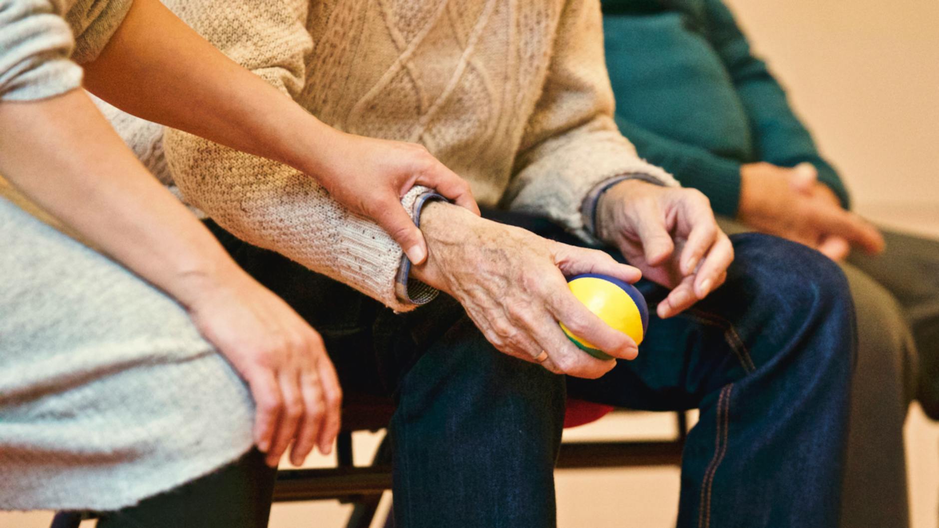
The correct diagnosis changes his treatment trajectory entirely, moving from stretching and ibuprofen to disease-modifying biologic medications that can slow or halt the inflammatory destruction of his joints. For families dealing with dementia, this scenario may seem remote, but inflammatory arthritis in a caregiver or in a patient with early-onset dementia can profoundly affect functional capacity and quality of life if left undiagnosed. Infection, while far less common, is another cause spine specialists must rule out. Bacterial infection of the bone, known as osteomyelitis or septic sacroiliitis, can directly involve the SI joint and cause inflammatory destruction. StatPearls documents this as a recognized though uncommon cause of SI joint dysfunction. Patients who are immunocompromised, including older adults on immunosuppressive medications, face elevated risk. The key distinguishing feature is fever and rapidly worsening pain, which should prompt urgent evaluation rather than the wait-and-see approach appropriate for mechanical causes.

Biomechanical Factors Including Leg Length Discrepancy, Gait Changes, and Scoliosis
Three biomechanical causes deserve attention together because they share a common mechanism: asymmetric loading. Leg length discrepancy, whether true or apparent, creates uneven pelvic mechanics that overload one SI joint relative to the other. StatPearls recommends measuring leg lengths in all patients with suspected SI joint dysfunction, a simple clinical step that is nonetheless frequently skipped. Abnormal gait patterns, from any cause, produce a similar effect. The American Academy of Physical Medicine and Rehabilitation notes that any condition altering normal walking places increased repetitive stress on the SI joints and surrounding structures. Scoliosis rounds out this triad by creating chronic asymmetric weight distribution across the pelvis. The comparison worth making here is between correctable and non-correctable causes.
A leg length discrepancy of a centimeter or less can often be addressed with a heel lift, and the resulting improvement in SI joint symptoms can be dramatic relative to the simplicity of the intervention. An abnormal gait caused by knee osteoarthritis or a neurological condition may be partially correctable with physical therapy or assistive devices, but the underlying cause may not be fully reversible. Scoliosis in an older adult is typically structural and non-correctable, meaning management focuses on symptom control rather than biomechanical restoration. For older adults with cognitive decline, gait changes are particularly relevant. Dementia frequently alters walking patterns well before the person or their family notices the cognitive symptoms. Shorter stride length, wider base, and shuffling steps all redistribute forces through the pelvis in ways that can trigger or worsen SI joint dysfunction. Caregivers who notice new complaints of low back or buttock pain in someone with dementia should consider whether a gait change is contributing, because addressing the gait problem with physical therapy or appropriate walking aids may relieve the pain without medication.
Hypermobility, Obesity, and Why Some Patients Are More Vulnerable to SI Joint Dysfunction
Generalized ligamentous laxity and obesity represent two very different pathways to the same outcome: an SI joint that cannot adequately handle the loads placed upon it. Weill Cornell Neurosurgery describes hypermobility of the SI joint as a condition in which the ligaments connecting the sacrum to the pelvis are too loose, allowing excessive movement that causes pain and inflammation. This can be congenital, as seen in connective tissue disorders like Ehlers-Danlos syndrome, or acquired through injury, hormonal changes, or repeated microtrauma. The limitation to recognize with hypermobility-related SI joint dysfunction is that many standard treatments, including manipulation and mobilization, can actually make the problem worse by further destabilizing an already loose joint. Stabilization exercises and bracing are generally more appropriate first-line interventions. Obesity acts through a simpler mechanism: increased mechanical loading on the pelvis. Physiopedia notes that obese patients are more commonly affected by SI joint pain, which aligns with what spine specialists see across their patient panels.
However, obesity rarely acts alone. It typically amplifies other causes on this list, making degenerative changes progress faster, making post-surgical compensation more severe, and making gait abnormalities more mechanically punishing. The warning for clinicians and patients alike is that weight loss, while beneficial, should not be treated as a standalone solution. An obese patient with SI joint dysfunction caused by ankylosing spondylitis needs disease-specific treatment regardless of their weight. Approximately 25 percent of adult patients with chronic low back pain have the SI joint as a primary pain source, according to research published in PubMed. Among vulnerable populations, including older adults with multiple comorbidities, the percentage may be even higher because so many of the risk factors on this list, including degeneration, gait changes, prior surgery, and obesity, cluster in aging bodies. The practical takeaway is that SI joint dysfunction should be on the differential diagnosis for any older adult with persistent low back or buttock pain, particularly when lumbar imaging does not fully explain the symptoms.
How Repetitive Activity and Sports-Related Stress Contribute to SI Joint Dysfunction
Athletes and physically active individuals can develop SI joint dysfunction through repetitive microtrauma rather than a single acute injury. Expert Review of Neurotherapeutics identifies weight-bearing and torsional sports, including football, gymnastics, and golf, as common non-traumatic causes. The mechanism is cumulative: each repetition of a high-force movement through the pelvis creates minor stress at the SI joint, and over time the ligaments and cartilage degrade.
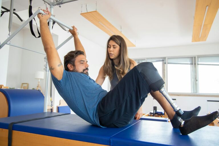
A recreational golfer who plays three times a week for twenty years may develop SI joint dysfunction not from any single swing but from tens of thousands of asymmetric rotational forces applied to the same joint. This cause is relevant to the aging population because many older adults maintain active lifestyles that include golf, swimming, or walking programs. These activities are generally excellent for overall health, but when SI joint pain develops, the specific movements involved need to be evaluated. Modifying technique, reducing volume, or temporarily substituting lower-impact activities can allow the joint to recover without abandoning physical activity entirely, a tradeoff that matters greatly for both musculoskeletal and cognitive health in older adults.
Where SI Joint Dysfunction Diagnosis and Treatment Are Heading
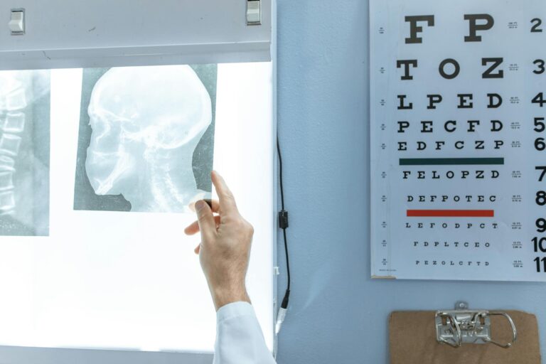
The diagnostic landscape for SI joint dysfunction is evolving. For decades, the condition was identified primarily through a combination of clinical provocation tests, with at least three positive tests out of five generally required to make the diagnosis. Diagnostic injections, in which anesthetic is placed directly into the SI joint under fluoroscopic guidance, remain the reference standard, but advances in MRI protocols are improving the ability to detect early inflammatory and degenerative changes non-invasively. For patients with dementia who may not be able to participate reliably in provocation testing, imaging-based diagnosis is becoming increasingly important.
Treatment has also progressed beyond repeated steroid injections. Minimally invasive SI joint fusion procedures have accumulated a growing evidence base over the past decade, offering a durable solution for patients who fail conservative management. For the broader dementia care community, the most important development may simply be increased awareness. As more primary care providers and geriatricians learn to include the SI joint in their evaluation of chronic low back pain, fewer older adults will endure prolonged courses of ineffective treatment directed at the wrong anatomic structure.
Conclusion
SI joint dysfunction arises from a well-defined set of causes that spine specialists encounter regularly in clinical practice. Degenerative arthritis, traumatic injury, pregnancy-related hormonal changes, post-lumbar fusion biomechanical stress, ankylosing spondylitis, leg length discrepancy, abnormal gait, ligamentous laxity, obesity, infection, and scoliosis each contribute through distinct mechanisms, but they share a common endpoint: pain and functional limitation that can significantly impair quality of life. For older adults and those living with cognitive decline, untreated SI joint pain compounds existing challenges with mobility, balance, and daily function.
The most important next step for anyone experiencing persistent low back or buttock pain, especially pain that has not responded to treatment directed at the lumbar spine, is to ask a provider to specifically evaluate the SI joint. With accurate diagnosis, effective treatment options exist at every stage, from physical therapy and bracing to injection-based therapies and minimally invasive fusion. Addressing SI joint dysfunction is not just about relieving pain; for aging adults and those in the dementia care community, it is about preserving the mobility and independence that support both physical and cognitive well-being.
Frequently Asked Questions
How do I know if my low back pain is coming from my SI joint rather than my lumbar spine?
SI joint pain typically presents as a deep ache on one side of the lower back or buttock, often below the belt line, and may radiate down the back of the thigh. Unlike lumbar disc pain, it rarely extends below the knee. A spine specialist can use a series of provocation tests and, if needed, a diagnostic injection to confirm the source.
Can SI joint dysfunction cause sciatica-like symptoms?
Yes, SI joint dysfunction can mimic sciatica by producing pain that radiates into the buttock and posterior thigh. However, true sciatica involves compression of the sciatic nerve root, while SI joint-referred pain follows a different neurological pathway. Distinguishing between the two requires careful clinical examination.
Is SI joint dysfunction permanent?
Not necessarily. Many cases, particularly those caused by pregnancy, acute trauma, or correctable biomechanical factors like leg length discrepancy, resolve with appropriate treatment. Degenerative and inflammatory causes tend to be chronic but manageable. The key variable is accurate diagnosis and targeted intervention.
How common is SI joint dysfunction after spinal fusion surgery?
Clinical studies suggest that up to 75 percent of post-lumbar fusion patients develop SI joint degeneration within five years of surgery. Not all of these patients become symptomatic, but the rate is high enough that routine SI joint monitoring after fusion is warranted.
Does weight loss help SI joint pain?
Reducing excess body weight decreases mechanical loading on the SI joint and can improve symptoms, but weight loss alone is rarely sufficient if another underlying cause, such as degenerative arthritis or ankylosing spondylitis, is also present. It is best approached as one component of a comprehensive treatment plan.