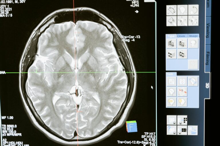
Gliosis on a brain scan refers to a process where the brain tissue shows signs of scarring or repair after injury. It happens when certain brain cells called glial cells multiply and form a dense network in response to damage. This reaction is the brain’s way of trying to protect and heal itself after events like trauma, stroke, infection, or other types of injury.
On imaging studies such as MRI, gliosis appears as areas that look different from normal brain tissue. These areas may show up as brighter or darker spots depending on the type of MRI sequence used. Gliosis is not a disease itself but a sign that the brain has undergone some form of injury or stress. It often accompanies other changes like loss of neurons or formation of cystic spaces where brain tissue has been lost.
Gliosis is important because it indicates that the brain has responded to damage, but it can also affect brain function depending on its location and extent. For example, gliosis in critical areas may contribute to neurological symptoms such as weakness, memory problems, or seizures. Doctors use brain scans showing gliosis to help understand the history of brain injury and guide treatment decisions.
In summary, gliosis is a scarring process in the brain visible on scans that marks areas where the brain has healed or reacted to injury. It reflects the brain’s attempt to repair itself but can also be linked to ongoing symptoms depending on the damage.
Sources
https://en.wikipedia.org/wiki/Creutzfeldt%E2%80%93Jakob_disease
https://pmc.ncbi.nlm.nih.gov/articles/PMC12571115/
https://www.braininjurylawofseattle.com/encephalomalacia-head-trauma/