Polyarteritis nodosa (PAN) is a rare and serious disease characterized by inflammation of medium and small arteries, which leads to damage in the blood vessel walls. This inflammation causes the arteries to weaken, narrow, or become blocked, resulting in reduced blood flow that can cause tissue injury or organ damage. The exact cause of polyarteritis nodosa remains unknown, but it is generally considered an autoimmune disorder where the body’s immune system mistakenly attacks its own blood vessels.
The underlying mechanism involves an abnormal immune response that triggers inflammation in arterial walls. In many cases, this may be related to hypersensitivity reactions—where the immune system overreacts to certain stimuli such as infections or other triggers—and leads to necrotizing arteritis (death of artery wall tissue). Immune complexes—combinations of antibodies bound to antigens—can deposit within vessel walls and activate complement proteins. This activation attracts white blood cells like neutrophils that release enzymes and reactive oxygen species damaging the vessel lining.
Some patients with PAN have been found to have associations with infections such as hepatitis B virus; this infection can stimulate chronic immune activation contributing to vascular injury. However, not all cases are linked with infections; many occur without any identifiable trigger.
The disease affects multiple organs because arteries supplying various tissues throughout the body are involved. Commonly affected sites include kidneys (leading to infarcts), gastrointestinal tract (causing abdominal pain), skin (resulting in rashes or ulcers), nerves (causing neuropathy), heart, and muscles. Symptoms often start nonspecifically with fever, weight loss, fatigue, muscle aches, and joint pains before more specific organ-related signs appear.
Histologically—the microscopic examination of affected vessels—shows segmental transmural inflammation involving all layers of artery walls along with fibrinoid necrosis where damaged tissue appears pinkish due to protein deposits resembling fibrin from clots.
While no single cause has been definitively identified for PAN:
– **Autoimmune factors** play a central role: The body’s immune system produces antibodies against components within its own vessels.
– **Immune complex deposition** contributes significantly: These complexes activate inflammatory pathways causing vessel wall destruction.
– **Infections**, particularly hepatitis B virus infection historically linked with some cases through persistent antigen stimulation.
– **Hypersensitivity reactions** may initiate or exacerbate vascular inflammation.
– Genetic predisposition might influence susceptibility but is not well defined yet.
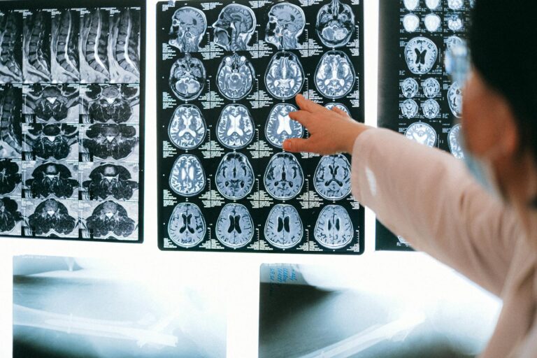
Because PAN involves systemic vasculitis affecting multiple organs variably among patients—with symptoms ranging from mild constitutional complaints like fever and malaise up to life-threatening organ ischemia—the diagnosis requires careful clinical evaluation supported by imaging studies showing characteristic arterial abnormalities such as aneurysms or stenoses (“beaded” appearance on angiograms).
Treatment focuses on suppressing this inappropriate immune activity using corticosteroids and immunosuppressive drugs which reduce inflammation and prevent further vascular damage. In cases associated with hepatitis B infection antiviral therapy alongside immunosuppression is necessary.
In summary, polyarteritis nodosa arises from an aberrant autoimmune attack on medium-sized arteries leading to necrotizing arteritis caused by complex interactions between hypersensitivity responses, immune complex deposition activating inflammatory cells that injure vessel walls resulting in multi-organ involvement manifesting diverse clinical symptoms depending on which arteries are affected most severely.