Alzheimer’s disease causes the brain to shrink progressively due to the loss of brain cells and the connections between them, a process known as cerebral atrophy. This shrinkage primarily affects critical areas involved in memory, thinking, and spatial awareness, such as the hippocampus and parts of the cerebral cortex. As neurons die and their networks break down, the brain tissue diminishes in volume, leading to enlarged fluid-filled spaces called ventricles and widened grooves on the brain surface.
At the core of this shrinkage are two abnormal protein accumulations: amyloid plaques and neurofibrillary tangles. Amyloid plaques are clumps of beta-amyloid protein that accumulate outside neurons, disrupting communication between cells. Neurofibrillary tangles are twisted fibers of tau protein found inside neurons, which interfere with the cell’s internal transport system and ultimately cause cell death. These protein abnormalities trigger a cascade of harmful effects, including inflammation and oxidative stress, which further damage neurons and accelerate brain tissue loss.
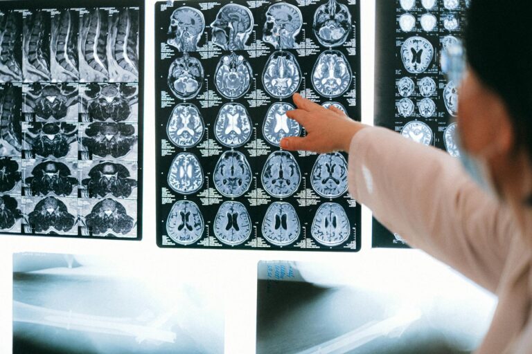
The hippocampus, a region essential for forming new memories, is among the earliest and most severely affected areas, leading to the hallmark memory problems in Alzheimer’s. Over time, the disease spreads to other parts of the cortex, causing difficulties with language, reasoning, and motor functions. This widespread neuronal loss results in the characteristic brain shrinkage visible on imaging scans such as MRI and CT.
Recent research suggests that other factors also contribute to brain shrinkage in Alzheimer’s. For example, abnormal accumulation of glycogen, a sugar stored in neurons, may disrupt energy management and increase oxidative stress, worsening neurodegeneration. Additionally, genetic risk factors like variations in the APOE gene influence susceptibility to Alzheimer’s and the extent of brain atrophy, although some mechanisms such as obesity-related brain injury appear to cause brain shrinkage independently of the classic amyloid and tau pathology.
The progression of brain shrinkage in Alzheimer’s can be divided into stages. Early on, subtle memory lapses correspond with mild hippocampal atrophy. As the disease advances, cortical thinning becomes more pronounced, and patients experience worsening cognitive and language impairments. In late stages, severe shrinkage affects multiple brain regions, leading to loss of motor control and complete dependence on caregivers.
Brain shrinkage in Alzheimer’s is not just a structural change but directly correlates with the clinical symptoms. The loss of neurons and synapses impairs the brain’s ability to transmit signals, causing memory loss, confusion, disorientation, and behavioral changes. Neurotransmitter levels also decline, further disrupting communication between brain cells.
Imaging techniques like MRI and PET scans are crucial for detecting brain shrinkage and the underlying protein deposits. MRI reveals structural changes such as hippocampal atrophy and ventricular enlargement, while PET scans can visualize amyloid and tau accumulation. Neuropsychological tests complement imaging by assessing cognitive decline and tracking disease progression.
In summary, the science behind brain shrinkage in Alzheimer’s disease involves a complex interplay of abnormal protein buildup, neuronal death, inflammation, oxidative stress, and metabolic disruptions. These processes lead to the progressive loss of brain tissue, especially in regions vital for memory and cognition, which underlies the devastating symptoms of the disease. Understanding these mechanisms is essential for developing treatments aimed at slowing or preventing brain atrophy and preserving cognitive function.