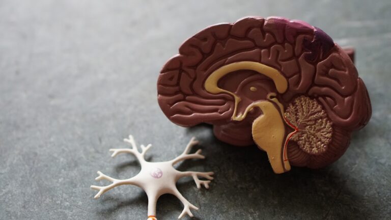
A brain MRI scan contains **no ionizing radiation at all**. Unlike X-rays or CT scans, which use ionizing radiation to create images, MRI (Magnetic Resonance Imaging) relies on strong magnetic fields and radiofrequency waves to generate detailed pictures of the brain and other soft tissues. This fundamental difference means that MRI scans do not expose patients to the radiation risks associated with ionizing radiation.
To understand why, it helps to know how MRI works. MRI machines use a powerful static magnetic field combined with radiofrequency pulses to excite hydrogen atoms in the body’s water and fat molecules. When these atoms return to their normal state, they emit signals that the MRI machine detects and converts into images. This process involves no ionizing radiation, which is the type of radiation that can remove tightly bound electrons from atoms and cause cellular damage. Instead, MRI uses non-ionizing radiation, which does not have enough energy to ionize atoms or molecules.
Because MRI does not use ionizing radiation, it avoids the risks linked to radiation exposure such as DNA damage, increased cancer risk, or tissue injury that can occur with X-rays or CT scans. This makes MRI a safer option for repeated imaging, especially in sensitive areas like the brain, and for vulnerable populations such as children or pregnant women.
However, MRI does have other safety considerations related to its strong magnetic fields. The magnetic field can attract ferromagnetic objects and interfere with implanted medical devices, so strict safety protocols are followed to screen patients and equipment before scanning.
In summary, a brain MRI scan involves **zero ionizing radiation**, making it fundamentally different and safer in terms of radiation exposure compared to other imaging methods like CT scans or X-rays. The images are produced through magnetic fields and radio waves, which do not carry the risks associated with radiation.