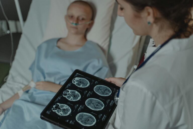
Detecting brain injury caused by asphyxia involves several specialized imaging tests that reveal the extent and location of damage to brain tissue due to oxygen deprivation. The most commonly used imaging techniques include magnetic resonance imaging (MRI), diffusion-weighted imaging (DWI), fluid-attenuated inversion recovery (FLAIR) MRI sequences, arterial spin-labeling (ASL) perfusion MRI, and cranial ultrasound. Each of these methods offers unique insights into the nature of brain injury from asphyxia.
**Magnetic Resonance Imaging (MRI)** is considered the gold standard for evaluating brain injury after asphyxia because it provides detailed images of soft tissues like the brain. MRI can detect areas where oxygen deprivation has caused cell death or swelling. It is particularly useful in identifying injuries in deep grey matter structures such as the thalamus, basal ganglia, hippocampus, and brainstem—regions highly vulnerable to hypoxic-ischemic damage. Specialized scoring systems applied to neonatal MRIs assess injury severity across different regions including white matter, cortex, cerebellum, and hemorrhages.
Within MRI techniques:
– **Diffusion-weighted Imaging (DWI)** measures water molecule movement within tissue; restricted diffusion indicates acute ischemic injury where cells are swollen or dying due to lack of oxygen. DWI is sensitive in detecting early changes following an anoxic event before they become visible on conventional MRI sequences.
– **Fluid-Attenuated Inversion Recovery (FLAIR)** sequences suppress fluid signals allowing better visualization of lesions near cerebrospinal fluid spaces; this helps identify edema or gliosis resulting from hypoxia.
– **Arterial Spin Labeling (ASL) Perfusion Imaging** noninvasively measures cerebral blood flow by magnetically labeling blood water molecules without contrast agents. ASL can detect abnormal perfusion patterns associated with anoxic injuries such as hyperperfusion or hypoperfusion in specific lobes which may correlate with clinical outcomes like seizures or loss of reflexes.
**Cranial Ultrasound** is often used initially in newborns because it is portable and safe at bedside but has limited resolution compared to MRI. It can show gross abnormalities such as bleeding or major structural changes but may miss subtle ischemic injuries typical after mild-to-moderate asphyxia.
Timing plays a crucial role: MRIs performed within days after rewarming from therapeutic hypothermia—a treatment used for infants suffering perinatal asphyxia—can reveal evolving patterns of injury that help predict neurodevelopmental outcomes later on.
In summary:
– Brain injuries from oxygen deprivation are best detected using advanced MRI techniques.
– DWI highlights acute cellular damage through restricted diffusion.
– FLAIR enhances lesion visibility near fluid spaces.
– ASL assesses cerebral blood flow abnormalities linked with prognosis.
– Cranial ultrasound serves mainly for initial screening but lacks sensitivity for detailed assessment.
These imaging tools together provide a comprehensive picture enabling clinicians to diagnose severity accurately, guide treatment decisions like therapeutic cooling timing, monitor progression over time, and counsel families about expected developmental challenges related to birth-related or other forms of asphyxial brain injury.