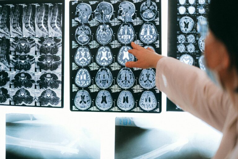
Magnetic Resonance Imaging (MRI) scans can detect signs related to inflammation in the brain, but the process is complex and indirect. MRI itself does not directly image inflammation as a specific entity; instead, it reveals changes in brain tissue and blood flow that often accompany inflammatory processes. These changes can include swelling, increased blood flow, breakdown of the blood-brain barrier, and alterations in brain structure or function that are associated with inflammation.
Inflammation in the brain involves activation of immune cells, release of inflammatory molecules, and changes in the brain’s microenvironment. When inflammation occurs, it can cause tissue swelling (edema), increased permeability of blood vessels, and sometimes damage to brain cells. MRI techniques are sensitive to these changes, which can be visualized as abnormalities in the images.
There are several MRI methods used to detect or infer brain inflammation:
1. **Conventional MRI**: Standard MRI sequences such as T1-weighted, T2-weighted, and FLAIR (Fluid-Attenuated Inversion Recovery) images can show areas of swelling or lesions that may be caused by inflammation. For example, in multiple sclerosis (MS), MRI reveals white matter lesions that reflect inflammatory demyelination. However, these images do not show inflammation directly but rather its consequences on brain tissue.
2. **Contrast-Enhanced MRI**: Using gadolinium-based contrast agents, MRI can highlight areas where the blood-brain barrier is disrupted—a hallmark of active inflammation. When the barrier is compromised, contrast leaks into brain tissue, making inflamed areas appear brighter on the scan. This technique is widely used in diseases like MS to identify active inflammatory lesions.
3. **Advanced MRI Techniques**:
– **Diffusion-weighted imaging (DWI)** and **diffusion tensor imaging (DTI)** can detect microstructural changes in brain tissue caused by inflammation.
– **Magnetic Resonance Spectroscopy (MRS)** measures chemical changes in brain tissue, such as increased levels of certain metabolites that may indicate inflammation.
– **Functional MRI (fMRI)** assesses changes in blood flow and oxygenation, which can be altered in inflammatory states, although fMRI is more commonly used to study brain activity than inflammation per se.
– **Quantitative MRI** methods, including volumetric analysis, can detect brain atrophy or tissue loss that may result from chronic inflammation.
4. **Emerging Techniques**: Research is ongoing into MRI methods that can more specifically target inflammation markers, such as imaging microglial activation (the brain’s immune cells) or detecting inflammatory proteins. These are not yet standard clinical tools but show promise for the future.
The ability of MRI to detect brain inflammation depends on the type, location, and stage of the inflammatory process. Acute inflammation with active blood-brain barrier breakdown is easier to detect with contrast-enhanced MRI. Chronic or low-grade inflammation may cause subtle changes that are harder to visualize.
In conditions like multiple sclerosis, MRI is a critical tool for detecting inflammatory lesions and monitoring disease activity. In other neurological disorders involving inflammation, such as neuropsychiatric lupus, encephalitis, or brain infections, MRI findings can support diagnosis by revealing patterns consistent with inflammation.
However, MRI findings must be interpreted carefully alongside clinical symptoms, laboratory tests, and sometimes other imaging modalities. MRI does not provide a definitive “inflammation” label but rather shows changes consistent with inflammatory processes. For example, swelling or lesions seen on MRI could also result from infection, ischemia, tumors, or other causes.
In summary, MRI scans are a powerful, non-invasive tool that can detect brain changes associated with inflammation, especially when using contrast agents and advanced imaging techniques. While MRI does not image inflammation directly, it reveals the structural and functional consequences of inflammation, aiding diagnosis and monitoring of neurological diseases involving inflammatory processes. Ongoing research aims to improve MRI’s specificity and sensitivity for detecting inflammation at the molecular and cellular levels withi