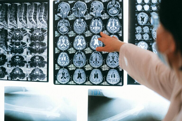
Brain scans can reveal a great deal about how Alzheimer’s disease progresses by showing changes in brain structure, function, and chemistry long before symptoms become obvious. These imaging techniques provide a window into the brain’s condition at different stages, helping doctors understand the disease’s development, monitor its advancement, and sometimes predict future decline.
Alzheimer’s disease typically unfolds gradually, starting with subtle brain changes that don’t immediately affect behavior or memory. This early phase, often called the preclinical stage, can last for years. Brain scans during this stage may detect abnormal accumulations of proteins like beta-amyloid plaques and tau tangles, which are hallmark features of Alzheimer’s. These proteins disrupt normal brain cell function and eventually lead to cell death. Specialized imaging methods such as PET (positron emission tomography) scans can visualize these protein buildups, revealing the disease’s silent beginnings even when a person feels cognitively normal.
As Alzheimer’s progresses into mild cognitive impairment (MCI), brain scans begin to show more noticeable changes. MRI (magnetic resonance imaging) scans can detect shrinkage in specific brain regions critical for memory and thinking, especially the hippocampus. This shrinkage reflects the loss of neurons and connections in these areas. Functional imaging techniques, like fMRI, can also reveal reduced activity or altered communication between brain regions involved in memory, attention, and problem-solving. These changes correlate with the subtle memory lapses and cognitive difficulties experienced during this stage.
When Alzheimer’s advances to dementia, brain scans typically show widespread brain atrophy, meaning significant loss of brain tissue across multiple regions. The ventricles, fluid-filled spaces within the brain, often enlarge as surrounding brain tissue shrinks. PET scans may continue to show extensive beta-amyloid and tau pathology. These imaging findings align with the more severe symptoms of dementia, such as profound memory loss, confusion, difficulty with language, and impaired daily functioning.
Beyond structural and protein imaging, brain scans can also assess metabolic activity. For example, FDG-PET scans measure glucose metabolism in the brain. Since neurons rely on glucose for energy, reduced glucose uptake in certain brain areas indicates impaired neuronal function. This metabolic decline often precedes visible atrophy and can help identify Alzheimer’s progression earlier.
Brain scans also play a role in differentiating Alzheimer’s from other types of dementia or brain disorders that may cause similar symptoms. By revealing specific patterns of brain changes and protein deposits, imaging helps ensure accurate diagnosis and appropriate treatment planning.
While brain scans provide critical insights, they are usually combined with clinical assessments, cognitive testing, and sometimes blood or cerebrospinal fluid tests to get a comprehensive picture of Alzheimer’s progression. Imaging alone cannot predict exactly how fast the disease will progress in an individual, but it significantly improves understanding of where the brain is along the disease continuum.
In recent years, advances in imaging technology and biomarker research have made it possible to detect Alzheimer’s-related changes earlier and more accurately than ever before. This early detection is crucial because it opens the door to interventions that might slow progression, improve quality of life, and help patients and families prepare for future challenges.
In essence, brain scans reveal the invisible story of Alzheimer’s unfolding inside the brain—from the earliest protein buildups and subtle functional shifts to the widespread damage and loss of brain tissue that underlie the disease’s devastating symptoms. They transform what was once a mysterious and hidden process into something visible and measurable, guiding diagnosis, research, and care.