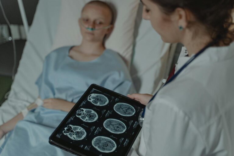
Electroencephalography (EEG) is a crucial tool used to monitor brain activity in babies who have experienced birth asphyxia, a condition where the newborn suffers from oxygen deprivation around the time of birth. This lack of oxygen can cause significant brain injury, and EEG helps doctors assess the extent of this injury, guide treatment decisions, and predict long-term neurological outcomes.
When a baby undergoes birth asphyxia, their brain cells may become damaged due to insufficient oxygen supply. EEG records electrical signals generated by neurons in the brain through electrodes placed on the scalp. In babies with birth asphyxia, continuous or amplitude-integrated EEG (aEEG) monitoring is often employed because it provides real-time information about brain function at the bedside in neonatal intensive care units.
The primary use of EEG in these cases is to detect abnormal background patterns that indicate how severely the brain has been affected. For example, an EEG might show suppressed or very low voltage activity if there is extensive damage. Conversely, more normal patterns suggest less severe injury. These background patterns are graded on scales developed specifically for neonates with hypoxic-ischemic encephalopathy (HIE), which results from birth asphyxia.
Another critical role of EEG monitoring is seizure detection. Seizures are common after birth asphyxia but can be subtle or even clinically silent in newborns; thus continuous EEG helps identify these events promptly so that appropriate anticonvulsant treatments can be started without delay.
In addition to traditional visual interpretation by neurophysiologists, advances such as machine learning algorithms are being developed to assist clinicians by automatically grading EEG background abnormalities and detecting seizures more accurately and efficiently within hours after birth.
EEG also plays an important part during therapeutic hypothermia treatment—a standard neuroprotective intervention for moderate to severe HIE—by allowing clinicians to monitor changes in cerebral function over time and adjust care accordingly.
Moreover, processed forms like amplitude-integrated EEG simplify complex raw data into trends that non-specialist staff can interpret easily at bedside for ongoing assessment during critical periods immediately following resuscitation from asphyxia.
Beyond seizure detection and severity grading, emerging research explores combining EEG data with other modalities such as near-infrared spectroscopy (NIRS) which measures cerebral oxygenation; together they provide insights into neurovascular coupling—the relationship between neuronal activity and blood flow—which may further refine prognosis prediction and guide interventions aimed at protecting vulnerable brains.
Overall, using EEG in babies affected by birth asphyxia enables early identification of those at highest risk for adverse outcomes including developmental delays or cerebral palsy. It informs clinical decisions regarding respiratory support needs, medication administration including anticonvulsants or sedatives during cooling therapy protocols, timing for advanced imaging studies like MRI scans done days later for structural assessment—and long-term follow-up planning focused on neurodevelopmental rehabilitation services when necessary.
In practice:
– Electrodes are carefully placed on specific scalp locations adapted for neonates.
– Continuous recordings start soon after delivery if perinatal distress suggests possible HIE.
– The recorded signals reveal whether cortical electrical activity is normal or shows suppression/attenuation.
– Seizure episodes appear as sudden bursts of rhythmic spikes distinct from baseline rhythms.
– Trends over hours/days help track recovery progress or deterioration.
This comprehensive monitoring approach allows multidisciplinary teams—including neonatologists neurologists nurses—to tailor supportive therapies precisely while minimizing secondary brain injury risks caused by uncontrolled seizures or inadequate oxygen delivery post-resuscitation.
Thus electroencephalography stands out not only diagnostically but also prognostically—it bridges immediate clinical management with longer-term outcome predictions—making it indispensable when caring for newborns suffering from hypoxic insults related to birth complications like prolonged labor cord compression placental insufficiency or emergency cesarean deliveries due to fetal distress signs detected via fetal heart rate monitors before delivery occurred.


