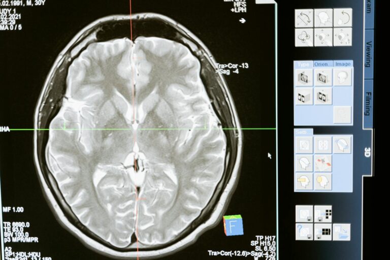
Doctors use a variety of sophisticated methods to limit collateral tissue damage during therapy, aiming to treat disease effectively while preserving as much healthy tissue as possible. These strategies are especially critical in treatments like radiation therapy, surgery, and certain drug therapies where the risk of harming normal cells is significant.
One key approach is **precision targeting**. In radiation therapy, for example, doctors use advanced imaging and delivery techniques to focus radiation beams very precisely on tumors, sparing surrounding healthy tissue. Techniques such as intensity-modulated radiation therapy (IMRT) and stereotactic body radiotherapy (SBRT) shape and modulate the radiation dose to conform tightly to the tumor’s shape. Additionally, temporary organ displacement methods physically move or shield nearby organs at risk during treatment, creating space between the tumor and healthy tissues to reduce exposure and damage.
Another important strategy involves **protective agents and biological enhancers**. Researchers have developed treatments that activate cellular defense pathways to reduce oxidative stress and inflammation caused by therapies like radiation. For instance, combining concentrated growth factors with specialized cell clusters can promote tissue regeneration, reduce harmful reactive oxygen species, and accelerate healing in damaged skin and soft tissues. These biological approaches help repair collateral damage and improve recovery after therapy.
**Hyperbaric oxygen therapy (HBOT)** is also used to limit tissue injury, especially in cases of radiation damage. By breathing pure oxygen under increased atmospheric pressure, patients increase oxygen delivery deep into tissues, which supports healing, reduces swelling, and stimulates new blood vessel growth. This enhanced oxygenation helps reverse tissue hypoxia (oxygen deprivation) caused by treatment and mobilizes stem cells that aid in tissue repair.
In surgical treatments, minimizing collateral damage involves using **precise, minimally invasive techniques**. For example, transoral laser microsurgery uses laser energy absorbed by water in tissues to cut or remove tumors with minimal heat spread, reducing injury to adjacent healthy structures. Surgeons also employ meticulous dissection methods and real-time imaging to avoid unnecessary trauma.
At the cellular level, therapies increasingly focus on **mitochondrial protection and modulation**. Since mitochondria play a central role in cell survival and energy metabolism, protecting them from therapy-induced dysfunction can reduce tissue injury. Experimental treatments include transferring mitochondria-rich vesicles to damaged tissues to restore energy balance and modulate immune responses, thereby improving healing and reducing inflammation.
Furthermore, the use of **computational modeling and digital twins**—virtual replicas of patient anatomy and physiology—allows clinicians to simulate and optimize treatment plans before actual therapy, predicting and minimizing collateral damage. Emerging materials and devices designed to shield or displace organs during treatment are also under development to enhance precision and safety.
In summary, doctors limit collateral tissue damage during therapy through a combination of precise targeting, protective biological agents, enhanced oxygen delivery, minimally invasive surgical techniques, cellular-level interventions, and advanced planning technologies. These integrated strategies help maximize treatment effectiveness while preserving healthy tissue function and promoting faster recovery.


