Doctors balance killing tumors with gamma rays against harming healthy tissue through a combination of precise targeting, careful dose planning, and advanced technology that maximizes radiation delivery to cancer cells while minimizing exposure to surrounding normal tissues.
Gamma rays are a form of ionizing radiation used in radiotherapy to destroy cancer cells by damaging their DNA, which prevents them from dividing and causes cell death. However, gamma rays can also harm healthy cells in the path of the beam, so the challenge is to deliver a lethal dose to the tumor while sparing as much normal tissue as possible.
To achieve this balance, several key strategies and technologies are employed:
**1. Precise Imaging and Tumor Localization**
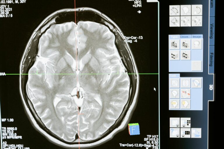
Before treatment, detailed imaging techniques such as CT scans, MRI, and PET scans are used to accurately locate the tumor and define its shape and size. This allows doctors to map out the exact area that needs to be targeted, ensuring that radiation is focused on the tumor volume and not unnecessarily spread to healthy tissue.
**2. Treatment Planning Systems (TPS)**
Using the imaging data, sophisticated computer software creates a three-dimensional model of the tumor and surrounding anatomy. The TPS calculates the optimal angles, intensities, and shapes of the gamma ray beams to conform the dose tightly around the tumor. This process is called dose planning or dosimetry. It involves balancing the dose high enough to kill tumor cells but low enough to protect normal tissue.
**3. Use of Multiple Beam Angles and Modulation**
Instead of a single beam, multiple gamma ray beams are directed at the tumor from different angles. Each beam individually delivers a lower dose, but where they intersect at the tumor, the doses add up to a high, tumoricidal level. This technique reduces the dose to healthy tissue along any single beam path.
**4. Advanced Delivery Technologies**
Devices like the Gamma Knife and linear accelerators (LINACs) are designed to deliver highly focused gamma rays with sub-millimeter precision. The Gamma Knife, for example, uses multiple cobalt-60 sources to converge gamma rays precisely on brain tumors, sparing surrounding brain tissue. LINACs can shape the radiation beam dynamically using multileaf collimators to conform to irregular tumor shapes.
**5. Fractionation of Dose**
Radiation is usually given in multiple small doses (fractions) over several days or weeks rather than one large dose. This approach allows normal cells time to repair some of the radiation damage between treatments, while cancer cells, which are less efficient at repair, accumulate lethal damage.
**6. Use of Monte Carlo Simulations and Quality Assurance**
Advanced computational methods like Monte Carlo simulations model how gamma rays interact with tissues, improving the accuracy of dose calculations. These simulations help validate treatment plans to ensure the prescribed dose distribution matches reality, reducing the risk of unintended damage.
**7. Consideration of Tumor and Tissue Radiosensitivity**
Different tumors and normal tissues vary in their sensitivity to radiation. Doctors consider these differences when planning treatment to optimize the therapeutic ratio—the balance between tumor control probability and normal tissue complication probability.
**8. Emerging Modalities and Particle Therapy**
While gamma rays are effective, newer forms of radiation such as proton and heavy ion therapy offer even more precise dose delivery with less collateral damage. These particles deposit most of their energy directly in the tumor with minimal exit dose, further protecting healthy tissue.
In essence, the balance is achieved by combining detailed imaging, precise computer planning, sophisticated beam delivery, and fractionated dosing to maximize tumor destruction while minimizing harm to normal tissue. This multidisciplinary approach requires careful coordination among radiation oncologists, medical physicists, dosimetrists, and radiotherapy technologists to tailor treatment to each patient’s unique anatomy and tumor characteristics.