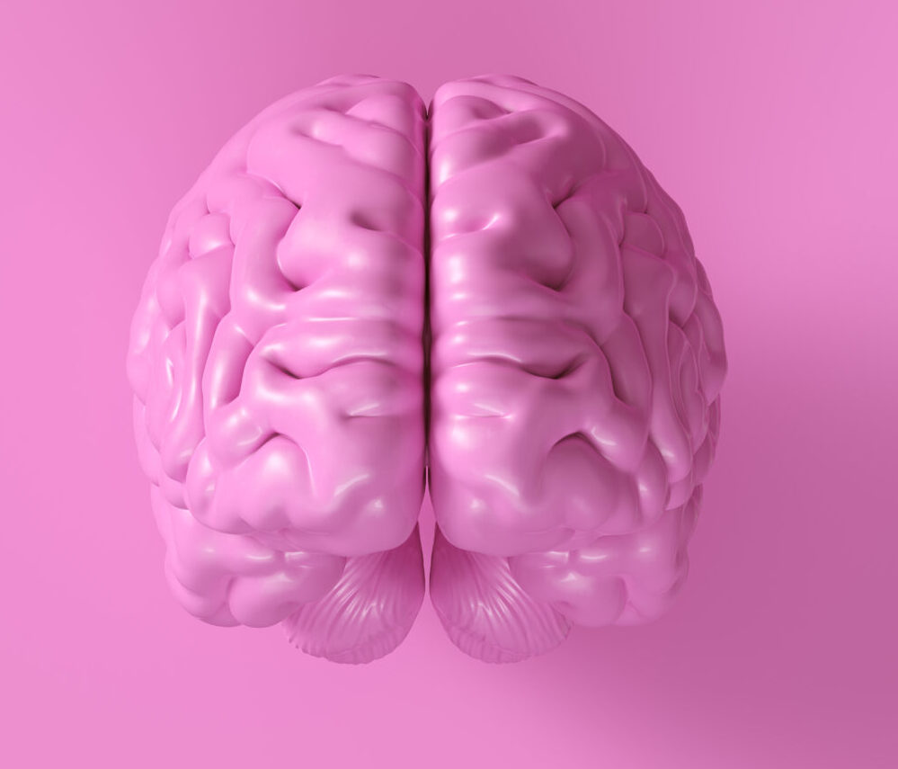
Advanced Neuroimaging to Map Alzheimer’s Progression
**Advanced Neuroimaging: Mapping Alzheimer’s Progression**
Alzheimer’s disease is a complex condition that affects the brain, leading to memory loss, cognitive decline, and other symptoms. To better understand and manage this disease, researchers are using advanced neuroimaging techniques to map its progression. Here’s how these technologies are helping us grasp Alzheimer’s better.
### 1. **Resting-State Functional Magnetic Resonance Imaging (rsfMRI)**
Resting-state functional magnetic resonance imaging (rsfMRI) is a method that looks at how different parts of the brain communicate with each other when a person is not actively doing anything. In Alzheimer’s disease, this communication can change over time. A recent study used rsfMRI to track these changes in people with Alzheimer’s and those at risk of developing it. The study found that in the early stages of Alzheimer’s, local brain activities decrease, while in later stages, long-range communication between brain regions is affected[1].
### 2. **Positron Emission Tomography (PET) Scanning**
Positron emission tomography (PET) scanning is another powerful tool for detecting Alzheimer’s. This method uses a radioactive tracer that binds to tau proteins, which accumulate in the brain as the disease progresses. Traditional PET scans have limitations in resolution, making it hard to detect tau deposition in small brain regions like the transentorhinal cortex. However, new high-resolution PET scanners, such as the PRISM-PET scanner, can detect tau accumulation even in these small areas. This technology is crucial for early detection and could make it possible to diagnose Alzheimer’s before symptoms appear[4].
### 3. **Electrophysiological Imaging**
Electrophysiological imaging involves recording electrical activity in the brain using techniques like local field potentials (LFPs) and electroencephalograms (EEGs). These methods help researchers understand how brain circuits change in Alzheimer’s disease. For instance, the Discrete Padé Transform (DPT) is a tool that can analyze multiple signals at once, providing insights into the complex dynamics of brain networks affected by Alzheimer’s. By studying these changes, scientists can identify early disturbances in brain circuitry, which could lead to better diagnostic tools and treatments[3].
### 4. **Biomarkers and Machine Learning**
Biomarkers are substances in the body that can indicate the presence of a disease. In Alzheimer’s, biomarkers like amyloid beta, tau proteins, and neurofilament light chain are being studied to predict the disease. Machine learning models, such as support vector machines (SVMs), can analyze these biomarkers to predict brain amyloidosis, which is a hallmark of Alzheimer’s. These models have shown promising results in different racial and ethnic groups, indicating their potential for personalized medicine[3].
### Conclusion
Advanced neuroimaging techniques are revolutionizing our understanding of Alzheimer’s disease. By mapping the progression of this complex condition, researchers can develop more accurate diagnostic tools and treatments. From resting-state fMRI to high-resolution PET scanning and electrophysiological imaging, these methods are providing a comprehensive view of how Alzheimer’s affects the brain. Additionally, biomarkers and machine learning models are helping predict the disease, paving the way for early intervention and better patient care. As research continues to advance, we can expect even more precise and effective strategies to manage Alzheimer’s.


