Assessing brain injury in newborns after asphyxia involves a comprehensive and multi-step approach that integrates clinical evaluation, neuroimaging, electrophysiological studies, and emerging biomarker technologies. The goal is to detect the extent and severity of brain damage early to guide treatment decisions and predict long-term outcomes.
First, clinical assessment begins immediately after birth with careful observation of the newborn’s neurological status. This includes evaluating muscle tone, reflexes, spontaneous movements, responsiveness to stimuli, breathing patterns, and consciousness level. Newborns who have experienced asphyxia often show signs such as lethargy or irritability, poor feeding ability, abnormal reflexes (like weak suck or Moro reflex), seizures, or respiratory difficulties. These signs help clinicians suspect hypoxic-ischemic encephalopathy (HIE), a common consequence of perinatal asphyxia.
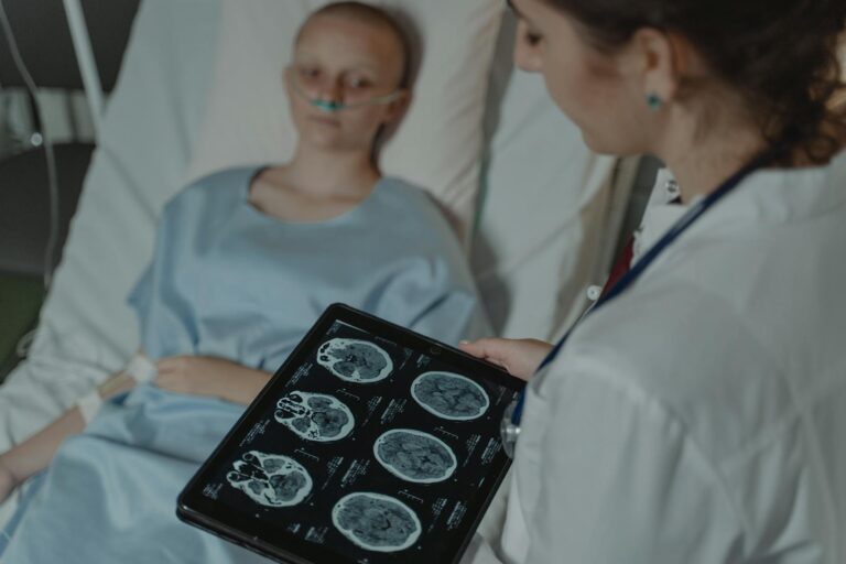
Next comes neuroimaging—one of the most critical tools for assessing brain injury in these infants. Magnetic Resonance Imaging (MRI) is considered the gold standard because it provides detailed images of brain structures without radiation exposure. Specifically:
– **Diffusion-weighted imaging (DWI)** MRI sequences are highly sensitive for detecting early ischemic changes within days after injury.
– The MRI scans are typically performed between 3 to 12 days after birth when the infant is stable enough for imaging.
– Radiologists use scoring systems like the Weeke score that systematically evaluate different brain regions including deep grey matter areas (thalamus and basal ganglia), white matter/cortex regions, cerebellum as well as hemorrhagic complications such as intraventricular hemorrhage.
– Injury severity in these areas correlates strongly with later neurodevelopmental outcomes.
In addition to MRI scans:
– **Cranial ultrasound** can be used at bedside soon after birth; although less detailed than MRI it helps identify gross abnormalities like bleeding or swelling quickly.
Electrophysiological monitoring plays a vital role too:
– **Electroencephalography (EEG)** records electrical activity from the newborn’s brain over time.
– EEG helps detect seizures which may be subtle clinically but harmful if untreated.
– Patterns on EEG can also indicate overall brain function impairment severity.
Biomarkers are an emerging frontier in this field:
– Researchers study blood-based markers released by injured neurons or glial cells shortly after hypoxia.
– Placental pathology examination may provide clues about prenatal insults contributing to neonatal injury risk.
Advanced techniques under development include artificial intelligence applications that analyze multimodal data—combining imaging features with EEG patterns and biomarkers—to improve accuracy in predicting outcomes.
Finally, ongoing neurological examinations during hospitalization track recovery progress or deterioration. Motor assessments help identify early signs of cerebral palsy risk due to specific regional injuries detected on imaging.
All these methods together form a layered approach: starting from bedside clinical observations through sophisticated imaging and electrophysiology studies supplemented by novel molecular insights—all aimed at timely diagnosis enabling targeted interventions such as therapeutic hypothermia which improves survival and reduces disability if started promptly within six hours post-injury.
This comprehensive assessment framework continues evolving rapidly thanks to advances in technology allowing earlier detection with greater precision while opening doors for personalized therapies tailored according to each infant’s unique pattern of injury.