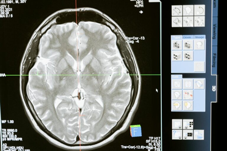
Yes, radioactive isotopes can be detected inside the body using specialized medical imaging techniques. These isotopes, often called radiotracers or radiopharmaceuticals, are introduced into the body and emit radiation that can be captured by sensitive detectors to create images or provide information about physiological processes.
When a radioactive isotope is administered—usually by injection—it travels through the bloodstream and accumulates in specific organs or tissues depending on its chemical properties. As these isotopes decay, they emit particles or photons such as gamma rays or positrons. Medical imaging devices detect this emitted radiation externally without invasive procedures.
One common method is **positron emission tomography (PET)**. In PET scans, a positron-emitting isotope undergoes decay inside the body; when a positron meets an electron nearby, they annihilate each other producing two gamma photons traveling in opposite directions. Detectors arranged around the patient capture these photons simultaneously to pinpoint where in the body the isotope is concentrated. This allows doctors to visualize metabolic activity and detect abnormalities like tumors because cancer cells often take up more of certain tracers than normal tissue.
Another widely used technique is **single-photon emission computerized tomography (SPECT)** which detects gamma rays emitted directly from radioactive tracers injected into patients. SPECT cameras rotate around the patient’s body capturing multiple images from different angles that a computer reconstructs into detailed 3D pictures showing how blood flows through organs such as heart or brain.
**Technetium-99m** is one of the most commonly used radioactive isotopes for diagnostic purposes because it emits gamma rays suitable for detection but has a short half-life minimizing radiation exposure to patients. It can be attached to molecules targeting specific tissues—for example, heart muscle cells during myocardial perfusion imaging—to assess blood flow and function under stress conditions.
These nuclear medicine techniques have several advantages:
– They provide functional information about how organs work rather than just structural details.
– They allow early detection of diseases before anatomical changes become visible on traditional X-rays or CT scans.
– The amount of radioactivity used is carefully controlled to balance image quality with patient safety.
However, there are some limitations:
– The spatial resolution—the ability to see very small structures—is lower compared with high-resolution CT or MRI.
– Movement during scanning can blur images.
– Radiation exposure exists but modern protocols minimize risks significantly.
In summary, detecting radioactive isotopes inside the human body relies on their natural emission of detectable radiation after administration as tracers targeted toward specific biological processes. Imaging technologies like PET and SPECT capture this signal externally providing valuable insights into health conditions ranging from cancer diagnosis to cardiac function assessment without invasive surgery.


