Doctors use gamma rays in radiotherapy primarily to treat cancer by targeting and destroying cancerous cells while minimizing damage to surrounding healthy tissue. Gamma rays are a form of high-energy electromagnetic radiation capable of penetrating deep into the body, making them effective for reaching tumors located inside organs or tissues. In radiotherapy, these rays are carefully controlled and directed to deliver a lethal dose of radiation to cancer cells, causing damage to their DNA and preventing them from growing or dividing.
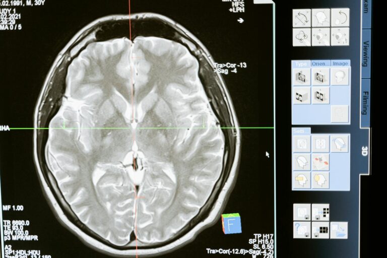
One of the most advanced applications of gamma rays in radiotherapy is through a technology called the Gamma Knife. This device is specifically designed for treating brain tumors and certain brain disorders without the need for invasive surgery. The Gamma Knife uses multiple focused beams of gamma radiation that converge precisely on the tumor site. Each individual beam is relatively weak and harmless to the healthy tissue it passes through, but where all beams meet, the combined radiation dose is high enough to destroy the tumor cells. This precision allows treatment of tumors that are difficult or risky to operate on, such as those near critical brain structures, with minimal side effects. The accuracy of the Gamma Knife is within half a millimeter, which significantly reduces radiation exposure to normal brain tissue and often requires only a single treatment session.
In more general external beam radiotherapy, gamma rays are produced by machines like linear accelerators (LINACs) that generate high-energy photon beams. These beams are shaped and modulated to conform to the tumor’s size and shape, sparing as much healthy tissue as possible. Treatment planning systems use sophisticated computer simulations to calculate the optimal dose distribution, ensuring the tumor receives enough radiation to be effective while protecting nearby organs. The dose is delivered in multiple sessions over several weeks to allow healthy cells time to recover between treatments.
Gamma rays kill cancer cells mainly by damaging their DNA. When gamma radiation passes through cells, it ionizes molecules and breaks DNA strands. This damage can be direct or indirect, the latter occurring through the creation of reactive oxygen species that further harm cellular components. Cancer cells, which divide rapidly and have less efficient repair mechanisms, are more vulnerable to this damage than normal cells. When the DNA damage is too severe, the cancer cells undergo programmed cell death or lose the ability to reproduce, leading to tumor shrinkage.
Doctors carefully calculate the radiation dose to balance effectiveness and safety. Too little radiation may fail to control the tumor, while too much can cause severe side effects by harming healthy tissue. Advances in technology now allow real-time monitoring of DNA damage during treatment, enabling personalized adjustments to radiation doses for better outcomes.
Besides brain tumors, gamma ray radiotherapy is used to treat various cancers, including head and neck tumors, lung cancer, prostate cancer, and others. It can also treat non-cancerous conditions such as arteriovenous malformations and certain functional brain disorders like trigeminal neuralgia.
In some cases, internal radiation therapy (brachytherapy) complements external gamma ray treatments. This involves placing radioactive sources close to or inside the tumor, delivering a high dose locally with minimal exposure to surrounding tissues.
Overall, gamma rays in radiotherapy represent a powerful tool in modern cancer treatment. Their ability to penetrate deeply and be precisely focused allows doctors to target tumors effectively, improving survival rates and quality of life for many patients. The continuous development of technologies like the Gamma Knife and advanced linear accelerators, combined with improved imaging and dose calculation methods, keeps enhancing the safety and success of gamma ray radiotherapy.