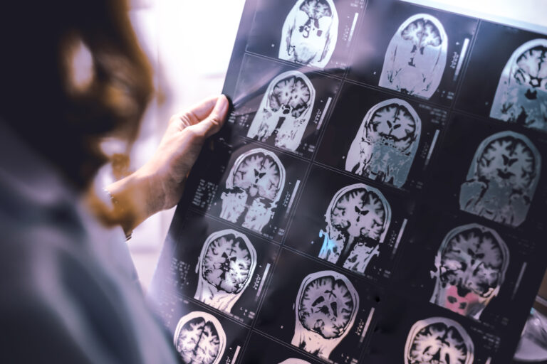
A head CT scan for concussion typically involves exposure to ionizing radiation in the range of about **2 to 4 millisieverts (mSv)**. This amount is roughly equivalent to a few months’ worth of natural background radiation that people receive from the environment annually. The exact dose can vary depending on the CT machine, scanning protocol, and whether contrast dye is used, but brain CT scans generally use lower doses compared to other types like abdominal or pelvic CTs.
To put this into perspective, natural background radiation exposes an average person to about 3 mSv per year. A single brain CT scan delivers a dose comparable to several months of this natural exposure. For example, if you consider that a chest X-ray gives around 0.1 mSv (about 10 days of background radiation), then a head CT’s dose is significantly higher but still relatively low in absolute terms.
The reason why a head CT scan uses more radiation than an X-ray is because it produces detailed cross-sectional images by taking multiple X-ray measurements from different angles and reconstructing them into slices of the brain tissue. This requires more energy and thus results in higher radiation doses.
Radiation doses are carefully controlled during medical imaging procedures with modern scanners designed to minimize exposure while still providing clear diagnostic images necessary for evaluating conditions like concussion or traumatic brain injury. Radiologists follow safety principles such as ALARA (“As Low As Reasonably Achievable”) which means they use the smallest possible amount of radiation needed for accurate diagnosis.
While there is some concern about cumulative effects if multiple scans are done over time—especially in children who are more sensitive—the risk from one single head CT scan remains very small relative to its clinical benefits when diagnosing serious conditions after head trauma.
In summary:
– A typical **head CT scan delivers approximately 2–4 mSv**.
– This dose equals several months’ worth of normal environmental background radiation.
– It’s much higher than simple X-rays but necessary for detailed brain imaging.
– Modern technology aims at minimizing exposure without compromising image quality.
– The lifetime cancer risk increase from one scan is very low but should be considered if repeated scans occur frequently.
– Children and pregnant women require special consideration due to increased sensitivity or potential fetal risks; alternative imaging methods like MRI or ultrasound may be preferred when appropriate.
Understanding these facts helps patients weigh the benefits and risks when undergoing a head CT after concussion symptoms appear, ensuring informed decisions guided by healthcare professionals who prioritize safety alongside diagnostic accuracy.


