Brain scans can provide valuable information about changes in brain structure and function that may be associated with dementia. However, it’s important to note that brain scans alone are not sufficient for diagnosing or predicting dementia.
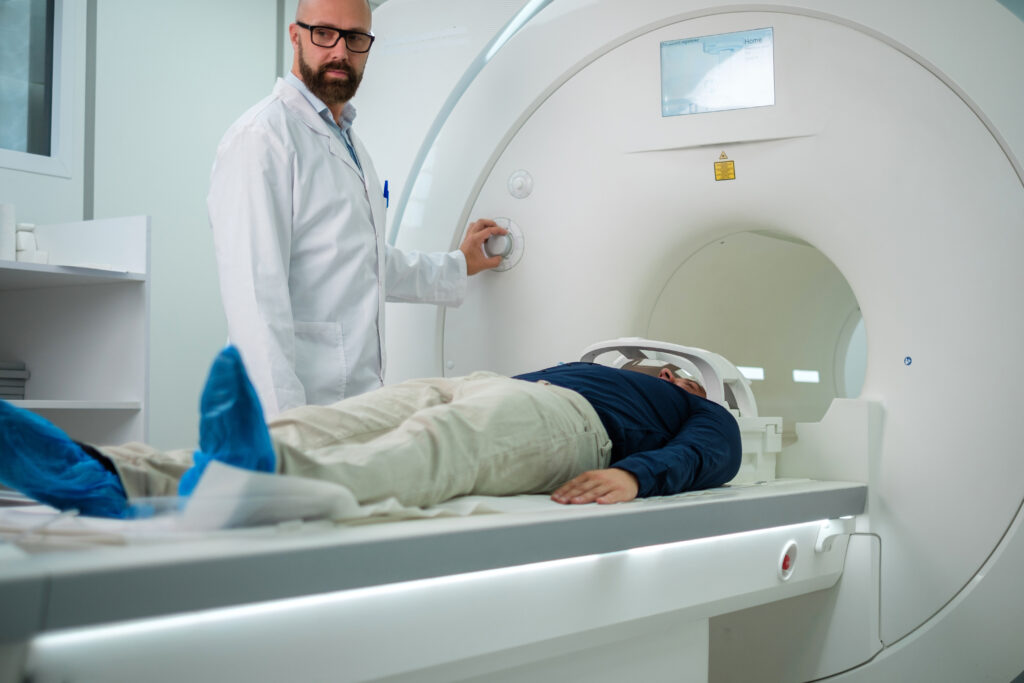
There are several types of brain scans that can be used to assess the brain in individuals with dementia, including magnetic resonance imaging (MRI), computed tomography (CT), and positron emission tomography (PET) scans. These scans can help identify areas of the brain that may be affected by dementia, such as areas of reduced brain volume, changes in brain connectivity, or the presence of abnormal protein deposits.
While brain scans can be useful in detecting these changes in the brain, they are not definitive for diagnosing dementia. A diagnosis of dementia typically requires a comprehensive evaluation, which may include a medical history, cognitive and neuropsychological assessments, and laboratory tests.
Furthermore, brain scans may not be able to predict the future development of dementia in individuals who do not yet show symptoms. While some studies have shown that changes in brain structure and function can be detected years before the onset of clinical symptoms, it is still not possible to predict with certainty which individuals will develop dementia.
There are several types of brain scans, and each has its own unique features and uses. Here are some of the most common types:
- Magnetic Resonance Imaging (MRI): MRI uses a powerful magnetic field and radio waves to produce detailed images of the brain. It is a non-invasive scan that can show structural changes in the brain, such as areas of reduced brain volume or the presence of lesions.
- Computed Tomography (CT): CT scans use X-rays to produce cross-sectional images of the brain. They are less detailed than MRI scans, but they can be useful for detecting areas of bleeding or swelling in the brain.
- Positron Emission Tomography (PET): PET scans use a small amount of radioactive material to show changes in brain function, such as changes in blood flow or the presence of abnormal protein deposits. PET scans are often used to help diagnose and monitor Alzheimer’s disease.
- Functional Magnetic Resonance Imaging (fMRI): fMRI is a type of MRI that measures changes in blood flow in the brain in response to different tasks or stimuli. It can be used to map areas of the brain that are involved in specific functions, such as language or movement.
Different types of brain scans have different strengths and weaknesses in detecting different types of dementia. Here are some general observations:
- Structural MRI scans are particularly good at detecting changes in brain structure, such as atrophy or lesions, and can be useful in diagnosing different types of dementia, including Alzheimer’s disease, Lewy body dementia, and frontotemporal dementia.
- PET scans that use a radiotracer that binds to beta-amyloid protein can help diagnose Alzheimer’s disease by detecting the presence of amyloid plaques in the brain. PET scans that use a radiotracer that binds to tau protein can also be used to help diagnose Alzheimer’s disease by detecting the presence of tau tangles in the brain.
- CT scans can be useful in detecting areas of bleeding or swelling in the brain, which may be a sign of vascular dementia.
- Functional MRI (fMRI) scans can be used to map areas of the brain that are involved in specific functions, such as memory or language, and can help identify patterns of brain activity that are associated with different types of dementia.
It’s important to note that brain scans alone are not sufficient for diagnosing dementia, and a comprehensive evaluation that includes a medical history, cognitive and neuropsychological assessments, and laboratory tests is needed. A diagnosis of Alzheimer’s disease, in particular, is typically made based on a combination of clinical symptoms, cognitive testing, and biomarkers, including brain imaging and cerebrospinal fluid analysis.
However, brain scans can provide valuable information that can help support a diagnosis of dementia and guide treatment decisions. In general, an MRI scan is the best initial test to evaluate the brain in individuals suspected of having dementia, but additional tests, including PET scans or fMRI scans, may be needed to confirm a diagnosis or help identify the specific type of dementia.
Can You Do an MRI Scan With a Pacemaker?
As for the question of whether these scans can be performed on individuals with pacemakers, it depends on the specific type of scan and the pacemaker. MRI scans are generally not recommended for individuals with pacemakers, as the strong magnetic fields can interfere with the pacemaker’s function. However, some newer pacemakers are designed to be safe for MRI scans. CT scans and PET scans are generally considered safe for individuals with pacemakers, but it is important to discuss any potential risks with your doctor.
Brain scans can provide valuable information about changes in brain structure and function that may be associated with dementia. However, it’s important to note that brain scans alone are not sufficient for diagnosing or predicting dementia.
There are several types of brain scans that can be used to assess the brain in individuals with dementia, including magnetic resonance imaging (MRI), computed tomography (CT), and positron emission tomography (PET) scans. These scans can help identify areas of the brain that may be affected by dementia, such as areas of reduced brain volume, changes in brain connectivity, or the presence of abnormal protein deposits.
Structural MRI scans are particularly good at detecting changes in brain structure, such as atrophy or lesions, and can be useful in diagnosing different types of dementia, including Alzheimer’s disease, Lewy body dementia, and frontotemporal dementia. However, MRI scans cannot definitively diagnose Alzheimer’s disease and cannot distinguish Alzheimer’s disease from other types of dementia.
PET scans can also be used to diagnose Alzheimer’s disease by detecting the presence of beta-amyloid plaques in the brain. PET scans that use a radiotracer that binds to tau protein can also be used to help diagnose Alzheimer’s disease by detecting the presence of tau tangles in the brain. PET scans are particularly useful for early diagnosis and can help distinguish Alzheimer’s disease from other types of dementia.
CT scans can be useful in detecting areas of bleeding or swelling in the brain, which may be a sign of vascular dementia. CT scans are less detailed than MRI scans but can still provide important information about changes in brain structure and function.
Functional MRI (fMRI) scans can be used to map areas of the brain that are involved in specific functions, such as memory or language, and can help identify patterns of brain activity that are associated with different types of dementia. fMRI scans are particularly useful for studying changes in brain function over time and can be used to monitor the progression of dementia.
It’s important to note that brain scans alone are not sufficient for diagnosing dementia, and a comprehensive evaluation that includes a medical history, cognitive and neuropsychological assessments, and laboratory tests is needed. A diagnosis of Alzheimer’s disease, in particular, is typically made based on a combination of clinical symptoms, cognitive testing, and biomarkers, including brain imaging and cerebrospinal fluid analysis.
In conclusion, brain scans can provide valuable information that can help support a diagnosis of dementia and guide treatment decisions. In general, an MRI scan is the best initial test to evaluate the brain in individuals suspected of having dementia, but additional tests, including PET scans or fMRI scans, may be needed to confirm a diagnosis or help identify the specific type of dementia. It’s important to work with a healthcare professional who specializes in dementia care and to undergo a thorough evaluation to ensure an accurate diagnosis and appropriate treatment plan.
References:
- Ossenkoppele, R., Schonhaut, D. R., Schöll, M., Lockhart, S. N., Ayakta, N., Baker, S. L., … & Rabinovici, G. D. (2016). Tau PET patterns mirror clinical and neuroanatomical variability in Alzheimer’s disease. Brain, 139(5), 1551-1567.
- Petersen, R. C., Caracciolo, B., Brayne, C., Gauthier, S., Jelic, V., & Fratiglioni, L. (2014). Mild cognitive impairment: a concept in evolution. Journal of Internal Medicine, 275(3), 214-228.
- Pinner, M. J., & Bouybayoune, I. (2019). Tau pathology in Alzheimer disease and Down syndrome
- “Brain Imaging: The Chemistry of Mental Activity” by Henry N. Wagner Jr. – This book provides an overview of the chemical and metabolic processes involved in brain activity and how they can be studied using imaging techniques such as PET and MRI.
- “The Clinical Neurobiology of the Hippocampus: An Integrative View” by Henriette van Praag and Fred H. Gage – This book explores the role of the hippocampus in memory and emotion, and how it can be studied using neuroimaging techniques.
- “MRI in Practice” by Catherine Westbrook and John Talbot – This book is a comprehensive guide to MRI imaging and its clinical applications. It covers basic principles of MRI, imaging techniques, and interpretation of MRI scans for various conditions, including brain disorders.